Helpline :
9807 55 6789
- Call Us Now: 9807 55 6789Call Us Now: 9807 55 6789
A comprehensive guide to PRP therapy — what it is, why concentration matters, the science behind the sting, and what the latest research genuinely says.
Let me start with something honest. When patients come to me asking about PRP — sometimes after reading about it in a newspaper, sometimes after their neighbour told them it fixed their knee — there is often a mixture of hope and scepticism in the same sentence. That is entirely understandable. PRP has been both overpromised and unfairly dismissed over the past two decades. Neither extreme serves you well.
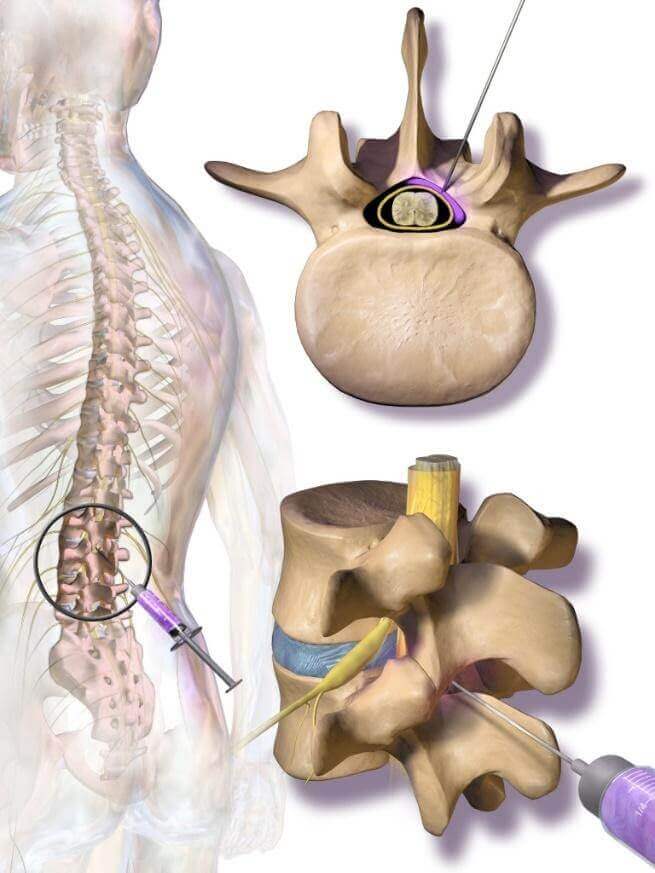
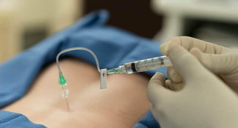
Platelet Rich Plasma, or PRP, is simply a concentration of your own blood's platelets. We draw a small volume of blood — typically 20 to 60 ml, not very different from a standard blood test — spin it in a centrifuge to separate its layers, and collect the fraction richest in platelets. This is then injected into the area of pain or damage. That is the procedure, mechanically speaking. But the biology is considerably more interesting.
Your blood contains red cells, white cells, plasma, and platelets. Platelets are small, disc-shaped fragments whose primary job in nature is to stop bleeding — they rush to a wound, clump together, and form a clot. But that is only half their story. Platelets are also warehouses. Packed inside them are hundreds of bioactive molecules — growth factors, cytokines, chemokines — that are released the moment a platelet becomes activated. These molecules speak directly to the cells around them, telling them to divide, migrate, differentiate, and rebuild.
PRP does not heal tissue directly. It is a signalling agent — a biological foreman that coordinates the cells already present in your tissue, waking up resident stem cells and amplifying the body's own repair machinery. The platelet is the messenger; your own cells do the actual rebuilding.
The key growth factors released include Platelet-Derived Growth Factor (PDGF), Transforming Growth Factor-β (TGF-β), Vascular Endothelial Growth Factor (VEGF), Insulin-Like Growth Factor (IGF), Epidermal Growth Factor (EGF), and Fibroblast Growth Factor (FGF). Each plays a different role — some promote collagen synthesis, some stimulate new blood vessel formation, some suppress inflammatory cascades, some recruit stem cells. Together, they create an environment that shifts damaged, degenerating tissue from a state of chronic non-healing inflammation into active repair.
This is where a lot of confusion lives. Patients hear "PRP" and assume there is one standard thing. There isn't. The type of PRP prepared depends on the centrifuge protocol, the collection system, whether white blood cells are included or removed, and whether a fibrin scaffold is created. Each variant has different biological properties and is suited to different clinical situations.
In leucocyte-poor PRP, the white blood cells (leucocytes) are deliberately removed during preparation. The result is a preparation that is high in platelets and growth factors but low in pro-inflammatory mediators. This is the preferred formulation for intra-articular (inside-the-joint) injections — particularly the knee, hip, and shoulder — because joint cartilage is an environment where you want regeneration without triggering excessive synovial inflammation. Several well-designed trials, including a landmark 2021 Cochrane-adjacent systematic review, found LP-PRP superior to corticosteroids for knee osteoarthritis at six and twelve months, with a more durable effect.
Leucocyte-rich PRP retains the white blood cells in the preparation. This creates a more inflammatory milieu upon injection — which sounds counterintuitive for a pain treatment, but makes biological sense for tendons and ligaments. Tendons are notoriously hypovascular — they have poor blood supply, which is exactly why tendon injuries heal so poorly. The controlled inflammation triggered by LR-PRP helps recruit cells to an otherwise quiet tissue and jump-starts a healing response that would not otherwise occur. LR-PRP is therefore the preferred choice in conditions such as lateral epicondylitis (tennis elbow), plantar fasciitis, patellar tendinopathy, and rotator cuff partial tears.
This is the newer, and in many respects the most sophisticated, member of the family. iPRF is prepared using a lower centrifuge speed, which keeps the platelets and leucocytes together in a fibrin network — a biological gel scaffold. Think of it as PRP with a slow-release mechanism built in. Instead of releasing all growth factors immediately upon injection, the fibrin matrix continues releasing them over days to weeks. The concentration of growth factors in iPRF is often higher than in conventional PRP, and the sustained delivery may explain some of the more durable results now being reported in facial aesthetics, dentistry, and increasingly in musculoskeletal medicine. At IBAP, we selectively use iPRF in combination protocols where sustained biological signalling adds value beyond what conventional PRP achieves.
Think of conventional PRP as a fire alarm that goes off once, loudly, and then stops. Think of iPRF as a slow-burning log on a fire — the biological signal continues building warmth in the tissue for weeks rather than minutes. Both have their place. The question is whether you need an acute signal or a sustained one.
This is arguably the most important thing I can tell you about PRP. And it is the reason why so many patients had PRP treatments years ago, saw no benefit, and concluded that PRP does not work. They were right that their treatment did not work. They were wrong that PRP itself doesn't work. The problem was dosing.
Your baseline platelet count in whole blood is roughly 150,000 to 350,000 platelets per microlitre. A "PRP" preparation that delivers only 2× baseline — perhaps 400,000 to 600,000 per microlitre — simply does not contain enough growth factor payload to produce a meaningful tissue response. Early commercial PRP kits, particularly those used in the late 2000s and early 2010s, frequently delivered these kinds of disappointing concentrations. The randomised trials that found PRP ineffective in that era were largely using these low-concentration preparations.
The emerging consensus in regenerative medicine, supported by in-vitro studies and increasingly by clinical dosing studies, is that a therapeutically effective PRP preparation needs to deliver approximately 5 to 7 times baseline platelet concentration — roughly 1 to 2.5 million platelets per microlitre in the final preparation. When calculated across a standard injection volume, this corresponds to approximately 10 billion platelets in a single injection.
There is also a ceiling to this relationship. Very high concentrations — above 8 to 10× baseline — appear, in some laboratory studies, to become inhibitory rather than stimulatory. It is a dose-response curve that plateaus and then turns down, not a simple "more is better" relationship. This is why modern PRP protocols are precision-calibrated, not simply "the more platelets the better." At our clinics, we use validated preparation systems with documented concentration outputs, not generic centrifuge programmes.
The early scepticism about PRP was earned, not manufactured. Studies from 2009–2015 using first-generation systems showed inconsistent results because inconsistent concentrations produced inconsistent biology. The subsequent meta-analyses that dismissed PRP were pooling studies of inadequately dosed preparations. The field has moved on considerably. Judging modern high-concentration, leuco-differentiated PRP by those old trials is like dismissing modern laparoscopic surgery because early procedures in the 1970s had high complication rates.
I am frequently asked: "Doctor, how many injections will I need?" The honest answer, backed by the best available evidence, is that a course of three injections — spaced four to six weeks apart — consistently outperforms a single injection in terms of both the degree of improvement and the duration of benefit.
The reasoning is biological. A single PRP injection triggers a healing response. But cartilage repair, collagen remodelling, and tendon regeneration are slow processes that occur over months. A second and third injection, timed at the point when the first cycle of growth factor signalling is winding down, sustains the biological momentum. Think of it like irrigation cycles for a damaged field: one good rain helps, but repeated seasonal rains allow the soil to recover fully and grow back strong.
The 2022 multi-centre trial by Filardo et al. and subsequent network meta-analyses have consistently shown that patients receiving three injections at monthly intervals report superior VAS (visual analogue pain scale) and WOMAC (functional outcome) scores at twelve months compared to those receiving a single injection. The difference is not marginal — it is clinically meaningful. Some protocols for knee osteoarthritis extend to four injections in patients with Kellgren-Lawrence grade III changes, where tissue repair needs extended biological scaffolding.
That said: some patients, particularly those with early-stage pathology, respond excellently to a single well-dosed injection. The protocol should be individualised, not templated. At IBAP, we assess the response after the first injection before finalising the course — if the patient reports a clear improvement, we continue; if the response is absent, we reassess whether the diagnosis, the technique, or the preparation may need adjustment.
One thing I always tell patients before their PRP injection: you may feel worse before you feel better. And this is not a failure. It is, in most cases, evidence that the treatment is doing exactly what it is supposed to do.
When PRP is injected and platelets are activated, they release their growth factor payload within minutes. This triggers an acute inflammatory response in the surrounding tissue — the same cellular cascade that occurs when tissue is freshly injured. In biological terms, you are creating a controlled, supervised version of an injury, to trick a chronically degenerate tissue into behaving like it has just been hurt and needs to heal.
For three to five days after injection, it is entirely normal — and actually desirable — to experience increased pain at the injection site, warmth, swelling, and occasionally a flu-like fatigue. Patients often ring us on day two asking if something has gone wrong. It hasn't. We reassure them, and by day seven, most are not only back to baseline but often beginning to notice the early green shoots of improvement.
If inflammation persists beyond 10–14 days, if the area becomes hot, red, and tracking (spreading redness), if you develop fever, or if the pain becomes severe and escalating rather than improving — please contact us immediately. While uncommon, infection at an injection site requires prompt assessment. Prolonged inflammation beyond two weeks may also indicate an exaggerated immune response that warrants evaluation.
I also want to address something that I see in clinic — patients who have been told by well-meaning family members to "rest completely" after PRP. Gentle, pain-guided movement is actually beneficial in the days after injection, as controlled loading helps align the new collagen fibres being laid down. Complete immobilisation is not recommended. We provide specific post-injection guidance at every visit.
PRP does not have to work alone. In fact, some of the most impressive clinical results I have seen in my practice have come not from PRP in isolation, but from combining it with complementary biological agents or procedural techniques. This is where modern regenerative pain medicine becomes genuinely exciting.
Hyaluronic acid is a naturally occurring molecule in joint fluid that provides viscoelastic lubrication and has its own modest anti-inflammatory and chondroprotective properties. When combined with PRP, HA appears to extend the retention time of PRP in the joint space, maintain a moist biological environment that supports growth factor activity, and provide immediate symptomatic relief whilst the slower PRP-mediated repair is getting underway. We often use high-molecular-weight HA co-injected with or immediately following LP-PRP for knee and hip osteoarthritis. Evidence from multiple RCTs, including the 2021 Cochrane review update, suggests this combination is superior to either agent alone.
Bone Marrow Aspirate Concentrate brings something PRP alone cannot: mesenchymal stem cells. Derived from a small sample of bone marrow (typically the posterior iliac crest), BMAC contains pluripotent stem cells capable of differentiating into cartilage, bone, and tendon cells — not just signalling to existing cells but actually contributing to rebuilding the structural tissue. Combined PRP-BMAC protocols are particularly powerful in avascular necrosis (AVN), moderate-to-severe osteoarthritis, and complex ligament injuries. This is a more involved procedure, but for the right patient in the right clinical situation, the combination offers biological potency that neither preparation achieves individually.
Nanofat is prepared from micro-emulsified adipose (fat) tissue, rich in adipose-derived stem cells (ADSCs) and stromal vascular fraction. When combined with PRP, the dual stem cell and growth factor environment creates a sophisticated regenerative milieu. We have seen particularly encouraging results with combined PRP-Nanofat in degenerative joint conditions and certain soft tissue disorders. This is a relatively newer combination in musculoskeletal medicine, and the evidence base is still growing — but the early clinical signals are compelling.
Prolotherapy — the injection of hyperosmolar dextrose solution into ligamentous and tendinous attachment points — has been used since the 1950s to stimulate connective tissue repair. Modern Prolotherapy combined with PRP creates a layered regenerative protocol: Prolotherapy addresses the periligamentous and peritenodinous tissue whilst PRP targets the joint itself or the core tendon structure. The combination is particularly useful in hypermobility-related pain and in conditions where ligamentous laxity is a driving factor.
This is an important conceptual point that I often need to explain to patients. PRP is a regenerative treatment — it works over weeks to months, not days. In patients with severe pain who need immediate functional improvement to participate in rehabilitation or simply to sleep, we often pair PRP with Radiofrequency Ablation (or Cooled RF for the knee's genicular nerves) to provide immediate, reliable pain interruption while the biological repair is underway. Cooled RF ablation of the genicular nerves provides pain relief for 9 to 18 months in most patients with knee OA — and during that pain-free window, the simultaneously administered PRP can do its regenerative work without the patient being distracted by debilitating pain. This is not a compromise — it is actually elegant clinical logic.
| Condition | PRP Type | Evidence Quality | Key Finding | vs. Comparator |
|---|---|---|---|---|
| Knee Osteoarthritis | LP-PRP × 3 | High (RCTs + MA) | Superior VAS/WOMAC at 12 months vs. HA and saline | Better than corticosteroid at 6 months |
| Lateral Epicondylitis | LR-PRP × 1–2 | Moderate–High | Significant pain reduction; longer effect than corticosteroid | Superior at 6 months; CS wins short-term |
| Plantar Fasciitis | LR-PRP × 2–3 | Moderate | Reduction in plantar fascia thickness + pain | Comparable to CS; more durable at 12 weeks |
| Rotator Cuff Tears (partial) | LR-PRP × 2–3 | Moderate | Improved MRI healing + functional scores | Better than physiotherapy alone |
| Hip OA (early–moderate) | LP-PRP × 3 | Moderate | Clinically meaningful HOOS improvement at 6 months | Superior to HA single injection |
| Achilles Tendinopathy | LR-PRP × 2 | Low–Moderate | Mixed — some benefit in mid-portion tendinopathy | No clear superiority over eccentric loading alone |
| Patellar Tendinopathy | LR-PRP × 1–2 | Moderate | Better outcomes than dry needling at 6 months | Comparable to ESWT |
| Chronic Low Back Pain (facet) | LR-PRP × 2 | Emerging | Benefit in facet-mediated pain when combined with PRP intra-discal | Insufficient powered RCTs — promising pilot data |
The evidence is strongest for knee osteoarthritis and lateral epicondylitis — and it is on these conditions that I would feel most confident recommending PRP as a first-line regenerative option ahead of surgical discussion. The field is growing rapidly; trials that were unpowered in 2015 are being redesigned with proper concentration validation and PRP characterisation, and the 2024–2025 literature is considerably more optimistic than anything published a decade ago.
| Feature | LP-PRP | LR-PRP | iPRF |
|---|---|---|---|
| Leucocytes | Removed | Retained | Retained (low spin) |
| Inflammatory response | Low | High (therapeutic) | Moderate, sustained |
| GF release | Immediate, concentrated | Immediate, high | Sustained over days–weeks |
| Best for | Joint injection (knee, hip, shoulder) | Tendons, ligaments, fascia | Combination protocols, facial, dental, complex joint |
| Post-injection soreness | Mild | Moderate–significant (3–5 days) | Mild–moderate |
| Preparation time | 15–20 min | 15–20 min | 10–12 min (lower spin) |
| Evidence level (MSK) | High | Moderate–High | Emerging (growing rapidly) |
I would not be doing my job if I only told you the positives. PRP is not a magic cure. There are genuine, important limitations that every patient deserves to understand before committing to treatment.
PRP is contraindicated or requires significant caution in the following situations. This is not an exhaustive list — a full clinical assessment is always needed before any regenerative procedure.
| Category | Specific Contraindication | Reason |
|---|---|---|
| Haematological | Thrombocytopaenia (<100,000/µL), platelet dysfunction syndromes | Insufficient platelets to prepare effective PRP or risk of inadequate clotting |
| Oncological | Active malignancy, especially haematological | Growth factors may stimulate tumour cell proliferation |
| Infection | Active infection at or near injection site; systemic sepsis | Risk of seeding bacteria into the injection; impaired immune response |
| Medications | Anticoagulants (warfarin, DOACs) if unable to pause; NSAIDs within 7 days | Reduced platelet function; NSAIDs blunt the inflammatory signalling cascade |
| Autoimmune | Active inflammatory arthritis (RA, PsA, AS) — relative contraindication | Unpredictable immune response; may exacerbate synovitis |
| Pregnancy | Relative contraindication — insufficient safety data | Growth factor effects on foetal development unstudied |
| Anaemia | Haemoglobin <10 g/dL | Insufficient blood volume for safe draw; poor quality PRP likely |
| Recent corticosteroid | Systemic or local steroids within 4–6 weeks | Corticosteroids impair the platelet activation response that PRP depends on |
A personalised consultation with Dr Vijay Bhaskar will assess your condition, imaging, and medical history to determine whether PRP — and which type — is the most appropriate treatment for you.
2nd Floor, 284/A, Road No. 12,
above IDFC First Bank,
near Omega Hospitals, MLA Colony,
Banjara Hills, Hyderabad — 500034
Sy No. 2, 4th Floor, Plot No. 200,
beside South India Shopping Mall,
opp. Fortune Heights, Mythri Nagar,
Madeenaguda, Hyderabad — 500049
This article has been written by Dr Vijay Bhaskar Bandikatla for general informational and educational purposes only. It does not constitute medical advice, diagnosis, or treatment recommendation for any individual patient. PRP therapy — like all medical procedures — carries risks and benefits that must be assessed on an individual basis by a qualified clinician. The information presented reflects current evidence at time of publication and may change as new research emerges. PRP is not appropriate for every patient or every condition. Please consult a qualified pain specialist or appropriate medical professional before making any decisions about your healthcare. IBAP Clinics does not accept responsibility for decisions made solely on the basis of this article. Vijay Advanced Pain Clinics Pvt. Ltd. — Banjara Hills and Madeenaguda, Hyderabad.
EXCELLENTTrustindex verifies that the original source of the review is Google. Good pain relief with treatmentPosted on GoogleTrustindex verifies that the original source of the review is Google. Best for pain reliefPosted on GoogleTrustindex verifies that the original source of the review is Google. बहुत अच्छा लगा हमें ये ट्रिटमेंट करा के अच्छे डॉक्टर और अच्छे स्टाफ हैं और काम पैसे में अच्छा अच्छा ट्रिटमेंट होता हैPosted on GoogleTrustindex verifies that the original source of the review is Google. Very nice, good and helpful staf, overall good experiencePosted on GoogleTrustindex verifies that the original source of the review is Google. Very good doctors and good tritment Thank you doctorPosted on GoogleTrustindex verifies that the original source of the review is Google. One of the best clinic for the relief of back painPosted on GoogleTrustindex verifies that the original source of the review is Google. Very good treatment indo british clinic for every pain reliefPosted on GoogleTrustindex verifies that the original source of the review is Google. I have visited indo british pain clinic this is superb clinic for pain relief that's why I have given 5 start rating to this clinic ...Posted on GoogleTrustindex verifies that the original source of the review is Google. My name is Raja Sekhar Reddy Tummuri from kakinada city My father was suffering nervous pain from last 3 years … I have decided to join with this op … Now everything fine after treatment done from DR. vijay sir … Wonderful service received from INDO British advanced pain clinic Hospitals in Hyderabad @ Banjara Hills road no 12.. Each and every staff response , concern , support & everything it’s too good Thank you so much … Special thanks to Mr. DR. Vijay bhaskar Bandi Katla …👏🏻👏🏻👏🏻👏🏻🙏🙏Verified by TrustindexTrustindex verified badge is the Universal Symbol of Trust. Only the greatest companies can get the verified badge who has a review score above 4.5, based on customer reviews over the past 12 months. Read more

Founder & Interventional Pain Specialist — IBAP Clinics, Hyderabad
MBBS · DA · FRCA (London) · FFPMRCA (Pain Medicine, UK) · MBA (Hospital Management)
CCT (Anaesthesia & Pain Medicine, UK) · Advanced Pain Training (Cambridge University Hospitals)
DDSMed Sports Medicine (Chicago) · Fellowship in Neuromodulation & Advanced Pain (London)
Dr Vijay brings over 15 years of postgraduate training across the United Kingdom’s most prestigious institutions — including the Royal College of Anaesthetists, Cambridge University Hospitals, and a dedicated neuromodulation fellowship in London — to his practice in Hyderabad. He is one of very few clinicians in India trained to the level of FFPMRCA — the Faculty of Pain Medicine of the Royal College of Anaesthetists — the highest qualification in pain medicine available in the UK.
His specialist expertise spans the full spectrum of knee pain management: from precision PRP and BMAC injections to cooled radiofrequency genicular nerve ablation, intrathecal drug delivery, and spinal cord stimulation for refractory pain states. He manages cases ranging from the weekend cricketer’s torn meniscus to the elderly cardiac patient with end-stage OA who has been told there are no further options.
Home » Procedures » Platelet Rich Plasma (PRP) Therapy
In our pain clinic, we provide pain relief so you can regain your identity.
Banjara Hills
2nd Floor, 284/A, Road No. 12, above IDFC First Bank, near Omega hospitals, MLA Colony, Banjara Hills, Hyderabad, Telangana 500034.

Indo-British Advanced Pain Clinic © 2026. All Rights Reserved. Designed & Developed By Adroit
WhatsApp us