Helpline :
9807 55 6789
-
Call Us Now: 9807 55 6789Call Us Now: 9807 55 6789
A frank, evidence-based guide from the interventional pain specialist’s desk covering every layer of hip pain from early cartilage wear to bone necrosis, and the Indian life that quietly drives it all.
Key Takeaways
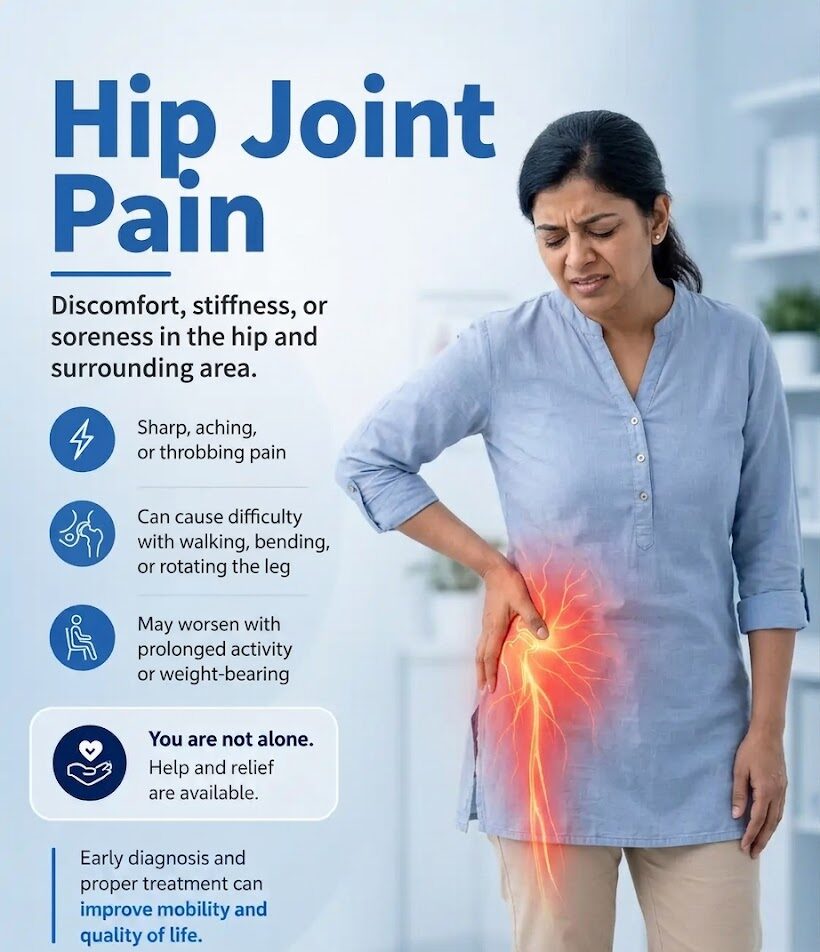
Hip pain is not one disease. It ranges from cartilage thinning (osteoarthritis) to bone death (avascular necrosis) to soft-tissue problems around the joint and much of it in India is driven by our unique lifestyle: long commutes over potholes, floor-level living, and hours of screen time. Regenerative treatments (PRP, BMAC, nanofat) are best in early-to-moderate stages. In AVN, Stage I–II is the window for joint preservation; Stage III–IV shifts to pain management and selective surgery. Cooled RFA provides 12–24 months of excellent analgesia for patients who cannot or will not have surgery. This is an honest guide not a sales pitch.
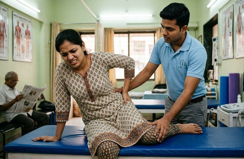
When a patient first tells me their hip hurts, I always pause before saying anything at all. Because “hip pain” in India is one of the most overloaded, under-diagnosed, and poorly treated conditions I encounter in my clinic. People have been limping for years. Told to “manage with painkillers.” Told “you need a replacement.” Or and this is equally unhelpful told “nothing is really wrong.”
The hip is a ball-and-socket joint. The ball is the head of the femur; the socket is the acetabulum. Between them sits articular cartilage a smooth, glistening surface about 2–4 mm thick that allows frictionless movement. When that cartilage degrades, you have osteoarthritis. When the blood supply to the femoral head is interrupted, you have avascular necrosis. When the tendons and bursae around the joint inflame, you have trochanteric bursitis or gluteal tendinopathy. And when the sacroiliac joint or piriformis muscle refer pain into the buttock and groin well, half the time we end up searching for a hip problem that was never really there to begin with.
Each of these has a different mechanism, different imaging findings, and a very different treatment algorithm. The commonest mistake I see even from well-meaning GPs and orthopaedic surgeons is treating all hip pain as one thing.
Think of the hip joint like a tyre. Osteoarthritis is slow, predictable tread wear you can see it coming and intervene early. Avascular necrosis is more like a sudden blowout from the inside the structure fails not because of overuse but because the rubber itself lost its supply of air. The treatment for each is completely different. You would not patch a blowout with the same repair kit you use for gradual tread wear.
Many of my patients arrive and say something like: “Doctor, I thought hip pain was only for very old people or after an accident. I just sit at a computer all day why am I suffering?” It is a fair question, and it deserves a real answer rather than a dismissive wave about age or stress.
Hip pain in urban India and particularly in a city like Hyderabad is a product of a very specific set of lifestyle stresses that we have somehow normalised. Let me be direct about what I see in clinic every week.
The software engineer who drives from Kondapur to Hitech City every morning 45 minutes each way, over speed breakers that could double as Olympic hurdles, white-knuckling every pothole on an ORR service road absorbs that impact through the spine, pelvis, and hip joint repeatedly. Every jolt transfers mechanical stress to the femoral head and the surrounding labrum. Then eight to ten hours of sitting at a workstation that is rarely set up correctly. Then the couch and the phone. Total daily hip movement: maybe 800 steps. Total daily hip compression: incalculable.
The homemaker who climbs stairs six times a day carrying full buckets of water, squats to mop with a floor-level jhadoo, sits on the kitchen floor to cut vegetables, and gets up and down from a low charpoy that is a different mechanical story, but the cumulative joint load is equally significant. Repeated deep squatting, in particular, drives the femoral head hard into the acetabulum at maximum range. Over years, the labrum frays. The cartilage thins at specific load zones.
Then there is the uncle who stands for ninety minutes on polished stone temple floors every Saturday and Sunday morning, waiting in the Balaji queue or attending a function, then sits in a cinema hall for three hours on inadequate cushioning. Stone floors transmit force without dampening. Prolonged standing on hard surfaces raises intra-articular hip pressure. It is not dramatic. It just adds up.
And teenagers yes, teenagers are increasingly presenting with hip discomfort. Coaching centre culture means eight to ten hours of sitting daily, often in chairs that are too high, bags that are too heavy, postures that no young spine should sustain. Early tendinopathy and labral strain are not rare in this group. They are just rarely looked for.
What I want you to understand is this: your pain has a cause. A structural, anatomical, identifiable cause. It is not just age. It is not just stress. It is not something to be endured and dismissed. And that cause whatever it turns out to be can almost always be addressed.
Before any treatment, we need to understand where the pain is actually coming from. I divide hip pain into three broad categories: intra-articular (from inside the joint), extra-articular (from surrounding soft tissues), and referred (from the spine, SI joint, or elsewhere). The location of your pain is the first clue.
Usually from inside the hip joint itself osteoarthritis, avascular necrosis (AVN), labral tear, femoroacetabular impingement (FAI). Often worsens putting on shoes or socks, getting in/out of a car, or crossing legs.
Usually from surrounding tendons and bursae greater trochanteric bursitis, gluteal tendinopathy, IT band irritation. Worst when lying on that side or climbing stairs sideways.
Often from the SI joint, piriformis, deep gluteal syndrome, or hamstring origin. May be provoked by sitting on hard surfaces think long auto-rickshaw rides or hard temple floors.
May not be hip at all more likely a “pinched nerve” from the lumbar spine or referred lumbar facet pain. Needs careful differentiation before any hip treatment is started.
The anatomical map above is a starting point not a diagnosis. What we do at IBAP Clinics is listen to the story carefully, examine gait, hip range of motion, pelvic alignment, and spine, and then confirm the pain generator with image-guided diagnostic injections. If we numb a specific structure and your pain disappears temporarily we know that is the culprit. This is precision medicine, not guesswork.
| 📍 Where Does It Hurt? | Likely Pain Source | Typical Triggers | Confirmed By |
|---|---|---|---|
|
🟢 Deep Groin /
Front of Hip |
Inside the hip joint — osteoarthritis, avascular necrosis (AVN), labral tear, FAI | Putting on shoes or socks, getting in/out of a car, climbing stairs, crossing legs | Intra-articular diagnostic injection under fluoroscopy |
|
🟠 Outer /
Lateral Hip |
Tendons and bursae — greater trochanteric bursitis, gluteal tendinopathy, IT band | Lying on that side at night, side-stepping, going up stairs | Ultrasound scan + bursa/tendon injection |
|
🟣 Buttock /
Posterior Hip |
SI joint, piriformis, deep gluteal syndrome, hamstring origin | Sitting on hard surfaces, long auto-rickshaw rides, temple floors, turning in bed | SI joint or piriformis diagnostic injection under imaging |
|
🟡 Pain Spreading
Down the Leg |
Often not the hip at all — lumbar disc, facet joint, or nerve root from the spine | Bending forward, coughing, prolonged sitting; often associated with back pain | Lumbar nerve or facet block; MRI of lumbar spine |
⚠ This table is a guide, not a diagnosis. Many patients have more than one pain generator. Overlap is common for example, hip OA and SI joint pain frequently coexist. Precise diagnosis always requires clinical examination and, where needed, image-guided diagnostic injection to confirm the true source before any treatment is planned.
Osteoarthritis (OA) of the hip is degenerative joint disease a slow, relentless thinning of the articular cartilage. The Kellgren–Lawrence (KL) scale grades it 0–4 on X-ray. In India, we are massively under-counting it, and a significant proportion of patients present at Grade 3 or 4 where options narrow considerably because Grade 1 and 2 pain was dismissed as “just age.”
Estimated hip & knee OA in India
Diagnosed only at moderate–severe stage
PRP vs HA at 6 months in KL 1–3 hip OA
Cooled RFA for advanced hip OA
I never jump straight to the most advanced treatment. Every patient deserves a logical, stepwise approach where each rung is tried appropriately before the next. That said, “stepwise” does not mean “slow” if a patient has significant pain and imaging supports an active biological treatment, I will recommend it without making someone suffer through weeks of inadequate analgesia first.


Think of your hip region like a busy Hyderabad traffic circle. Forces are entering from the spine, pelvis, thighs, and core from multiple directions. If one exit is blocked or overloaded say, a weak gluteal muscle you feel the “jam” as pain. But the actual cause may be at a completely different lane from where the honking is loudest. That is why we map the whole system before treating any single point.
Let me be direct here. Regenerative therapies PRP, BMAC, nanofat are not magic. They are biological treatments that harness the body’s own healing capacity. They work better when there is still meaningful cartilage and bone architecture present. Think of it like repainting a wall you can do a proper job if there is still plaster; but if the plaster has completely crumbled, you need to rebuild, not repaint.
PRP (Platelet-Rich Plasma) is prepared by centrifuging your own blood to concentrate growth factors PDGF, TGF-β, VEGF, IGF-1 that reduce inflammation and may stimulate chondrocyte activity. A 2023 systematic review of RCTs confirmed PRP outperforms hyaluronic acid in pain and function scores at 12 months for hip OA.
BMAC (Bone Marrow Aspirate Concentrate) is harvested from the patient’s iliac crest under local anaesthesia a 30-minute procedure. The concentrate is rich in mesenchymal stromal cells (MSCs), haematopoietic progenitors, VEGF, and anti-inflammatory cytokines. These components drive osteogenic and angiogenic repair exactly what is needed in ischaemic necrotic bone, and in articular cartilage that has begun to thin.
In patients with KL Grade 1 or 2 hip OA particularly younger patients we combine intra-articular (inside the joint) injections with fluoroscopy-guided femoral head injections of BMAC or PRP. These procedures can be performed together in a single session. The intra-articular component addresses the joint environment reducing inflammation, providing lubrication, and supporting the cartilage surface. The femoral head injection targets the subchondral bone and the early microvascular changes that precede advanced cartilage loss. Used together, the aim is to unlock the regeneration potential of the joint and postpone or entirely avoid surgery for as long as possible. This is not an experimental idea. It follows the same biological logic as regenerative protocols used in knee OA, adapted for the specific anatomy and demands of the hip.
Nanofat and SVF (Stromal Vascular Fraction) are mechanically processed fat emulsified and filtered to remove adipocytes while retaining pericytes, endothelial progenitors, and MSCs. The evidence base is thinner than for PRP or BMAC but growing rapidly. In selected patients, we mix BMAC or nanofat with PRP to maximise the growth factor payload delivered to the target tissue this is what I call a “biological cocktail” approach. The rationale is straightforward: BMAC brings the cells and the osteogenic drivers; PRP brings a concentrated burst of growth factors and anti-inflammatory cytokines; together, the two components work synergistically in a way neither achieves alone.
| Therapy | Best For | KL Stage | Evidence Level | Primary Goal | Duration |
|---|---|---|---|---|---|
| PRP intra-articular | Early–moderate OA | KL 1–3 |
Level I–II (RCTs) |
Analgesia + biological repair |
6–12 months |
| BMAC (intra-articular + femoral head) | KL 1–2 OA; AVN I–II | KL 1–2 | Level II–III |
Regeneration — postpone/avoid surgery |
9–18 months |
| PRP + BMAC biological cocktail | KL 1–2 OA; AVN I–II (young patients) | KL 1–2 |
Level II–III (emerging) |
Maximum regenerative payload |
9–18 months |
| Hyaluronic Acid (HAI) | Moderate OA, analgesia | KL 2–3 | Level I–II | Analgesia + lubrication | 3–6 months |
| Cooled RFA | Moderate–severe OA, inoperable | KL 3–4 | Level II–III | Analgesia only | 12–24 months |
| Prolotherapy | Ligament laxity, early OA | KL 1–2 | Level II–III | Structural support | 6–12 months |
| Corticosteroid | Acute flares | Any | Level I | Acute analgesia | 4–8 weeks |
Not every patient with Grade 4 hip OA is a surgical candidate. Some have cardiac or respiratory comorbidities that make anaesthesia genuinely dangerous. Some are elderly and simply do not want major surgery. Some are reluctant for personal reasons. And some a category I feel strongly about have had their pain dismissed with a shrug and a prescription.
These patients deserve active management, not passive abandonment.
Cooled RFA targets the articular sensory nerve branches supplying the hip joint — predominantly from the obturator nerve and the nerve to quadratus femoris. Internally cooled probes create larger, more consistent lesions than conventional RFA, ablating pain pathways without damaging motor function. A pilot study in Skeletal Radiology showed HOOS scores improving from 17 to over 52 at six months — allowing patients to resume daily activities and reduce opioid use. Recent retrospective data from 2024 confirms consistent results across academic pain centres, with repeat procedures showing maintained efficacy.
Imagine the hip joint is a building on fire. In late-stage OA, the building is genuinely damaged difficult to rebuild. But you can disconnect the fire alarm wire so it stops screaming continuously. The building still needs attention, but you the person inside can finally function. Cooled RFA doesn’t fix the joint; it silences the nerve that is broadcasting relentless pain signals.
AVN is, in many ways, the more tragic diagnosis. Whereas OA is a gradual wearing process, AVN is a vascular catastrophe. The femoral head depends on a delicate blood supply. When that supply is disrupted by corticosteroid use, alcohol, trauma, sickle cell disease, or sometimes for no identified reason bone cells die silently, often while the X-ray looks relatively normal. By the time pain is established and imaging shows collapse, significant structural damage may already have occurred.
In Hyderabad, I see AVN particularly in young adults in their twenties and thirties who received prolonged corticosteroids for nephrotic syndrome, rheumatoid disease, or COVID-related illness. If you are young and have unexplained groin pain, please request an MRI of the hip, not just an X-ray. MRI detects AVN months sometimes years before plain film changes appear.
The Ficat–Arlet system is the most widely used staging framework for AVN of the femoral head. I want to explain not just the staging but what we are actually trying to achieve at each stage because the goals of treatment shift fundamentally as disease progresses. In early stages, we are trying to preserve and regenerate. In late stages, we are trying to control pain and maintain quality of life.
An important clarification on technique: when I describe BMAC delivery into the femoral head for Stage I and II AVN, I am not necessarily referring to surgical core decompression in the traditional sense. At IBAP Clinics, we perform fluoroscopy-guided, fine-needle BMAC injection directly into the femoral head a minimally invasive, non-surgical procedure performed under live X-ray guidance as a day-case procedure. This allows precise delivery of the cellular concentrate into the necrotic zone without a formal orthopaedic operation, and is performed in combination with an intra-articular hip joint injection to provide both biological stimulation within the bone and a supportive biological environment within the joint itself. For patients who are surgical candidates, surgical core decompression combined with BMAC augmentation remains the gold standard with the strongest evidence and we will refer appropriately. But the non-surgical route offers a real and evidence-informed alternative, particularly for younger patients where the goal is to postpone or avoid surgery for as long as possible.
| Ficat Stage | Structural Findings | Imaging | 🌱 Regeneration Strategy | 💊 Analgesia Strategy | BMAC Suitability | Surgical Note |
|---|---|---|---|---|---|---|
|
Stage I
Pre-radiographic ischaemia
|
Normal X-ray. Blood supply compromised but bone not yet dead. | X-ray normal · MRI shows early signal change |
PRIMARY GOAL
PRP + fluoroscopy-guided BMAC injection directly into the femoral head (non-surgical, fine-needle technique under image guidance). Combined with intra-articular injection to unlock regeneration potential. Goal: neovascularisation & cell rescue — postponing or avoiding surgery for as long as possible.
|
SECONDARY
HAI for background pain. Oral analgesics short-term.
|
Highly Suitable Best prognosis. Small lesions respond well. |
Joint preserving. Non-surgical BMAC delivery preferred first; surgical core decompression referral if available and appropriate. |
|
Stage II
Structural damage without collapse
|
Cysts and sclerosis. Femoral head architecture intact. No crescent sign yet. | X-ray shows sclerosis/cysts · MRI shows necrotic area clearly |
PRIMARY GOAL
Non-surgical fluoroscopy-guided BMAC delivery into the femoral head — using a fine needle under image guidance, BMAC is delivered precisely into the necrotic zone without open surgery. This can be performed alongside intra-articular hip injection to unlock regeneration potential and maximise joint preservation. Combined with PRP as a biological cocktail. Evidence strongly supports this approach: superior Harris Hip Score and lower THA conversion vs core decompression alone.
|
SECONDARY
PRP injections ± HAI for pain between procedures.
|
Highly Suitable Strong meta-analysis support (954 subjects, 2025) |
Joint preserving. Non-surgical BMAC + intra-articular injection is the primary approach at IBAP. Surgical core decompression referral for appropriate candidates. |
|
Stage III
Subchondral collapse — crescent sign
|
Femoral head beginning to collapse. Mechanical integrity compromised. Crescent sign on X-ray. | X-ray: crescent sign · MRI: subchondral fracture |
LIMITED / DEBATED
BMAC may provide short-term symptomatic benefit but cannot reverse structural collapse. Lesion size (Kerboul angle / Kim & Koo index) matters — small lesions may still benefit. Discussed case-by-case.
|
PRIMARY GOAL
Cooled RFA of articular branches (obturator + femoral nerve). HAI for lubrication. PRP for biological analgesia. Oral analgesic optimisation (duloxetine, low-dose naltrexone).
|
Limited / Debated High THA conversion within 12–24 months. Useful only palliation in larger lesions. |
Assess surgical fitness. THA referral for suitable patients. For unfit patients — pain management is the primary strategy. |
|
Stage IV
Articular collapse + secondary OA
|
Femoral head collapsed. Secondary osteoarthritis of acetabulum. Joint space lost. | X-ray: OA changes, collapse · MRI: full structural failure |
NOT APPLICABLE
Regenerative therapies cannot address established structural failure at this stage.
|
PRIMARY GOAL
Cooled RFA for patients not fit for surgery. HAI, PRP for biological analgesia in selected cases. Full analgesic optimisation.
|
Not Indicated Structural failure beyond biological repair. |
Total hip arthroplasty (THA) is definitive treatment for surgical candidates. For those unfit or unwilling — comprehensive pain management programme. |
BMAC works in AVN because the necrotic femoral head has lost its native osteoprogenitor cell population it cannot repair itself. BMAC replenishes these cells along with VEGF and osteogenic growth factors, essentially resupplying what biology has depleted. But and this is critical once the femoral head has physically collapsed (Stage III crescent sign), no biological therapy can restore mechanical integrity. The evidence is consistent: smaller lesions at earlier stages respond far better. Lesion size (measured by the Kerboul angle or Kim & Koo index) matters as much as the Ficat stage itself.
Imagine the femoral head is a bridge whose foundations have been damaged by subsidence. In Stage I–II, the bridge structure is still standing intact but weakened. Core decompression drills relief channels to release pressure; BMAC injects repair mortar into those channels. Lorries (daily life) can keep crossing safely. In Stage III, the bridge deck has begun to buckle and crack. You can still make it passable carefully but you cannot restore its full load-bearing capacity with mortar alone. In Stage IV, the bridge has fallen. You build a new one (THA) or if that is not possible you reroute traffic around it (cooled RFA, analgesic management).
A significant proportion of patients I see with “hip pain” actually have pain originating from structures around the joint, not within it. The distinction matters enormously because the treatment is completely different. Getting this wrong means treating the wrong target and wondering why the patient is not improving.
The greater trochanteric bursa sits between the greater trochanter and the iliotibial band and gluteal tendons. When inflamed from altered gait, leg length discrepancy, obesity, or repetitive stair climbing it causes lateral hip pain worst when lying on that side at night. In Hyderabad, I see this particularly in middle-aged women who stand for long periods on hard kitchen floors and in office workers who then compensate with an evening walk that stresses the IT band further. Treatment is stepwise: physiotherapy for hip abductor strengthening, ultrasound-guided corticosteroid injection as first step, PRP for refractory tendinopathic changes.
The piriformis is a small deep buttock muscle that can entrap the sciatic nerve, producing buttock pain with radiation down the posterior thigh mimicking disc-related sciatica. Sitting on hard surfaces for long periods is a key provocateur. Think long auto-rickshaw rides, hard cinema seats, stone temple floors, and the coaching-centre plastic chair that a teenager sits on for eight hours. Diagnosis is confirmed by ultrasound-guided diagnostic injection. Treatment includes physiotherapy, injection of local anaesthetic with corticosteroid or PRP, and occasionally botulinum toxin for truly refractory cases.
The SI joint connects the sacrum to the ilium and moves just 2–4 mm yet it is a powerful pain generator. The cultural practice of sitting on the floor for pooja, meals, social gatherings, cooking places the SI joint in extreme positions repeatedly across a lifetime. SI joint pain typically causes lower back, buttock, and posterior thigh pain, worst when rising from sitting, climbing stairs, or turning in bed. Treatment includes fluoroscopy-guided SI joint injections, prolotherapy to the SI ligaments for ligament laxity, and cooled or bipolar RFA of the lateral branches (L4–S3) for chronic refractory cases.
| Condition | Location | Key Provocative Feature | First-Line Treatment |
|---|---|---|---|
| Hip OA | Groin, anterior thigh | Walking, stairs, cross-legged sitting | PRP / HAI / Cooled RFA |
| AVN Femoral Head | Groin, medial thigh | Weight-bearing, night pain | Core decompression + BMAC (early) / CRFA (late) |
| Greater Trochanteric Bursitis | Lateral hip | Lying on side, side-step walking | US-guided CSI / PRP |
| Piriformis Syndrome | Deep buttock, posterior thigh | Sitting on hard surfaces, hip rotation | US-guided injection ± physio |
| SI Joint Pain | Lower back, buttock | Standing from sitting, turning in bed | Fluoroscopy-guided injection / RFA |
| Ischiogluteal Bursitis | Deep buttock at ischium | Sitting on hard surfaces | US-guided injection |
I want to say something that is not usually written in clinical articles. I say it because I mean it.
Hip pain is disabling in a way that is difficult to appreciate unless you have experienced it. It is not dramatic. It is slow, grinding, and relentless. It affects the most ordinary things lowering onto a toilet seat, getting in and out of a car, performing namaaz or attending a temple. It disrupts sleep, which then disrupts mood, concentration, and relationships. It makes people feel old before their time and makes them withdraw from activities that gave their life meaning.
In India, there is a cultural tendency to dismiss chronic pain as something to be endured stoically. “Everyone has some pain at this age.” This is not wisdom. It is abandonment normalised as resilience. When a 55-year-old man cannot walk to the local market, or a 45-year-old woman cannot sit through her son’s prize-giving without shifting painfully in her chair, that is not acceptable if something can be done about it.
Understanding a patient’s pain really understanding it, not just noting a score on a form is itself therapeutic. There is solid evidence from chronic pain research that patient-perceived empathy from their clinician independently improves pain outcomes, reduces analgesic requirements, and increases treatment adherence. The descending pain inhibitory pathways are modulated by psychological state, including the sense of being heard and valued. This is not soft science. This is neuroscience.
So when someone comes to IBAP Clinics and says “my hip has been hurting for three years and nobody has taken it seriously” I take it seriously. We investigate properly. We discuss honestly. And we plan treatment that is realistic, evidence-based, and tailored to the actual person in front of us.
Start your journey with a virtual consultation to discuss symptoms from home.
We review your medical history and relevant reports for a clear understanding.
Our doctors conduct a thorough assessment through detailed discussions.
We confirm findings with state-of-the-art imaging like X-rays, CT scans, and MRIs.
Our team identifies the root cause and key trigger points for treatment.
We create a customized treatment plan, including necessary medications and procedures.
Our Pain Specialists support a complete recovery focused on total wellness.
We provide ongoing follow-ups tailored to each treatment plan, ensuring continuous care and long-term recovery support.
We relieve your pain, helping you be yourself again!
Absolutely not. Hip pain arises from the joint itself (OA, AVN, labral tears, FAI), from surrounding soft tissues (bursitis, gluteal tendinopathy), or is referred from the spine or SI joint. Age is one risk factor but far from the only one. Young professionals, students, active homemakers, and two-wheeler riders all present with significant hip pain at IBAP Clinics — and most of them have a clear, addressable anatomical cause once we look properly.
Hip joint pain is typically felt in the groin and anterior thigh and worsens with walking, getting in/out of a car, and sitting cross-legged. Back-related referred pain tends to travel down the back of the thigh or into the lower leg. Many patients have both. A diagnostic injection under image guidance numbing the hip joint specifically is the gold standard for confirming whether the hip is the true pain generator.
Yes, when used appropriately. Image-guided corticosteroid injections are accurate, safe, and effective for acute flare-ups of hip OA or bursitis. They are not a long-term solution repeated steroid injections can potentially weaken cartilage and tendons over time. At IBAP Clinics, we use them as a bridge to more durable treatment, not as an end in themselves.
PRP (Platelet-Rich Plasma) is prepared from your own blood by centrifugation, concentrating growth factors PDGF, TGF-β, VEGF, IGF-1. Unlike corticosteroid injections which suppress inflammation temporarily, PRP works biologically to promote tissue repair, support chondrocyte activity, and modulate the inflammatory environment. Three-dose monthly protocols show better outcomes than single injections in hip OA.
Cooled radiofrequency ablation targeting the articular branches of the obturator and femoral nerves typically provides 12–24 months of meaningful pain relief. Some patients maintain benefit beyond two years. The procedure can be safely repeated if pain returns, and repeat sessions show consistent results. It does not treat the underlying arthritis but can dramatically improve quality of life in patients who are not surgical candidates.
In many cases, yes particularly in early-to-moderate OA (KL grades 1–3) and early AVN (Ficat stages I–II). Regenerative therapies, targeted injections, physiotherapy, and cooled RFA can provide long-lasting relief without surgery. Even in end-stage OA or late AVN where surgery is unsuitable, meaningful and sustained pain control is achievable through interventional pain management.
If your hip pain has persisted beyond 4–6 weeks, if physiotherapy and pain tablets have not provided adequate relief, if you want to explore non-surgical options, or if surgery carries significant risk given your health an interventional pain specialist can precisely map your pain generator and offer a structured treatment ladder from injections through regenerative therapies to RFA. We work alongside orthopaedic surgeons; a referral for surgery will always be made if and when it is the right answer for you.

EXCELLENTTrustindex verifies that the original source of the review is Google. Good pain relief with treatmentPosted on GoogleTrustindex verifies that the original source of the review is Google. Best for pain reliefPosted on GoogleTrustindex verifies that the original source of the review is Google. बहुत अच्छा लगा हमें ये ट्रिटमेंट करा के अच्छे डॉक्टर और अच्छे स्टाफ हैं और काम पैसे में अच्छा अच्छा ट्रिटमेंट होता हैPosted on GoogleTrustindex verifies that the original source of the review is Google. Very nice, good and helpful staf, overall good experiencePosted on GoogleTrustindex verifies that the original source of the review is Google. Very good doctors and good tritment Thank you doctorPosted on GoogleTrustindex verifies that the original source of the review is Google. One of the best clinic for the relief of back painPosted on GoogleTrustindex verifies that the original source of the review is Google. Very good treatment indo british clinic for every pain reliefPosted on GoogleTrustindex verifies that the original source of the review is Google. I have visited indo british pain clinic this is superb clinic for pain relief that's why I have given 5 start rating to this clinic ...Posted on GoogleTrustindex verifies that the original source of the review is Google. My name is Raja Sekhar Reddy Tummuri from kakinada city My father was suffering nervous pain from last 3 years … I have decided to join with this op … Now everything fine after treatment done from DR. vijay sir … Wonderful service received from INDO British advanced pain clinic Hospitals in Hyderabad @ Banjara Hills road no 12.. Each and every staff response , concern , support & everything it’s too good Thank you so much … Special thanks to Mr. DR. Vijay bhaskar Bandi Katla …👏🏻👏🏻👏🏻👏🏻🙏🙏Verified by TrustindexTrustindex verified badge is the Universal Symbol of Trust. Only the greatest companies can get the verified badge who has a review score above 4.5, based on customer reviews over the past 12 months. Read more
This article is intended for educational and informational purposes only. It does not constitute medical advice, diagnosis, or a treatment plan. All treatment decisions must be made in consultation with a qualified medical professional who can assess your individual clinical circumstances, imaging findings, and overall health. Regenerative therapies including PRP, BMAC, and nanofat are biological procedures whose evidence base is evolving; they are not universally approved for all indications. Results vary significantly between individuals and disease stages. IBAP Clinics and Dr Vijay Bhaskar Bandikatla accept no liability for actions taken based on the information in this article without professional clinical assessment.

MBBS, DA, FRCA (UK), FFPMRCA (Pain Medicine, RCOA, UK)
CCT (Anesthesiology And Pain Management)
Neuromodulation & Advanced Pain Research Fellowship (London), MBA (HM)
Banjara Hills
2nd Floor, 284/A, Road No. 12, above IDFC First Bank, near Omega hospitals, MLA Colony, Banjara Hills, Hyderabad, Telangana 500034.

Indo-British Advanced Pain Clinic © 2026. All Rights Reserved. Designed & Developed By Adroit
WhatsApp us