Helpline :
9807 55 6789
-
Call Us Now: 9807 55 6789Call Us Now: 9807 55 6789
From thermal RF to cryoablation — a plain-language guide to the technology that is quietly changing how chronic pain is managed in India.
Let me be honest with you. Pain is one of the most dismissed experiences in Indian healthcare. Patients come to me after years — sometimes decades — of being told "take this tablet and rest," or worse, "it is all in your head." It is not in your head. Pain is real, it is measurable, and now — thanks to the procedures I am going to describe here — it is often very treatable without surgery or a lifetime of medications.
Think about the average day in Hyderabad. You wake up, probably slept in an awkward position on a too-soft mattress — or a too-hard one. You drive to work, dodging potholes on Road No. 12, every speed-breaker sending a jolt through your spine. You sit for eight hours hunched over a screen. If you are in IT — and Hyderabad has tens of thousands of you — the hours blur into each other, the neck stiffens, the lower back screams quietly. You drive home. You watch television on a sofa that was never designed for your posture. And then you wonder why your back has been hurting for three years.
For middle-aged and older patients, I see another pattern. The temple visits, the long queue on those uneven stone floors, standing for forty minutes at Birla Mandir or Chilkur Balaji. Weekend films at the multiplex — three hours in a seat designed for someone with a shorter spine. None of these activities are harmful in themselves. But for someone who already has facet joint degeneration or sacroiliac joint dysfunction, they are the straw that keeps breaking the camel's back, week after week.
The point I am making — and I make this to every patient I see — is that chronic pain rarely has one cause. It is the accumulation of modern Indian life, falling on joints and nerves that were not designed to withstand it indefinitely. Understanding this context is, in my view, the first step towards better treatment. Empathy before prescription. Always.
Radiofrequency and cryo ablation procedures do not treat the underlying structural cause of pain — they interrupt the nerve pathway that carries the pain signal. This distinction matters enormously. Used correctly, these procedures can provide months or years of meaningful relief, allowing patients to rehabilitate, exercise, and reclaim their lives. They are tools in a broader strategy, not a permanent cure in isolation.
The nervous system is essentially a communication network — think of it like a telephone exchange. Pain signals are the calls. The nerve fibres are the telephone wires, and your brain is the exchange that processes the incoming calls and makes you aware of pain. Ablation procedures — whether using heat, cold, or pulsed energy — work by temporarily or semi-permanently cutting those wires, so the pain signal simply cannot complete its journey to your brain.
There are four principal technologies used in modern interventional pain medicine. Each has distinct physics, distinct clinical applications, distinct equipment, and a different duration of effect. Understanding the differences will help you ask better questions — of your doctor, and frankly, of any clinic that offers these procedures.
Imagine your pain nerve is a CCTV cable sending a live feed of suffering to your brain's control room. Thermal RFA simply cuts the cable — clean, effective, but the cable will eventually regrow. Cooled RFA cuts a wider section of cable — more reliable disruption. Pulsed RF scrambles the signal without cutting the cable at all — reversible, safer near sensitive wires. Cryoablation freezes the cable until it shatters — it will regrow, but along the same route, giving you time before the feed returns.
Thermal RFA is the original — and still the most widely used — ablation technique in pain medicine. It works on the principle of resistive heating. A radiofrequency generator delivers an alternating current at approximately 500 kHz through an insulated needle to an exposed metal tip. The tissues immediately surrounding that tip resist the flow of electrical current, and this resistance generates heat — reaching 80 to 90 degrees Celsius. At this temperature, the small nerve fibres carrying pain signals (specifically the C-fibres and Aδ-fibres) are irreversibly coagulated. The lesion produced is roughly cylindrical, about 5 to 7 mm in diameter.
The procedure is performed under fluoroscopy (live X-ray guidance) or ultrasound. Before the actual ablation, we perform sensory and motor nerve testing at the electrode tip — this is critical. We want to confirm we are at the correct nerve before heating, and equally confirm we are not at a motor nerve that controls movement.
Thermal RFA is the gold standard for lumbar and cervical facet joint denervation — treating the medial branch nerves that supply the facet joints. These are the small joints that run along your spine, and they are a primary source of back and neck pain in a large proportion of patients. It is also used for thoracic facet pain, and increasingly for the genicular nerves of the knee (the nerves supplying the arthritic knee joint) in patients who are not yet ready for or not suitable for joint replacement.
Onset is typically 2 to 4 weeks after the procedure, as post-procedural inflammation settles. Some patients notice improvement within days; others take a month. Duration of effect is 12 to 24 months. When the nerve regrows — and it does, slowly — the procedure can be safely repeated, often with similar or better results.
You need a dedicated radiofrequency generator (Stryker, Abbott/St. Jude, Cosman are established brands), specifically designed, insulated cannulae with different active tip lengths, a fluoroscopy or C-arm unit, and a trained interventional pain physician. This is emphatically not a procedure for a general practitioner or a physiotherapy clinic — the mapping, testing, and real-time guidance are what separate a good outcome from a complication.
Now this is where it gets interesting — and where many clinics in India fall behind, because cooled RFA requires more sophisticated equipment and training. In conventional thermal RFA, the highest temperature is right at the electrode tip. This creates a relatively small lesion, which works well when the target nerve has a predictable, consistent anatomical location (as with lumbar medial branch nerves).
But what about the sacroiliac joint? The lateral branch nerves supplying the SI joint are notoriously variable in their location — from patient to patient, even from left side to right side in the same person. A standard thermal RFA electrode might miss the target nerve entirely. Cooled RFA solves this elegantly. An internal pump circulates water through the electrode tip, preventing the tip itself from overheating and charring the tissue. This allows radiofrequency energy to penetrate further outward, creating a much larger, spherical lesion — 8 to 12 mm in diameter. Think of it as the difference between a focused spotlight and a floodlight. The floodlight covers more area.
Cooled RFA is the preferred technique for sacroiliac joint pain — one of the most under-diagnosed causes of lower back pain, particularly in women after pregnancy and in patients following a fall or road traffic accident. It is also used for genicular nerve ablation in osteoarthritic knees (under the COOLIEF® brand), hip joint denervation, and shoulder pain. In Hyderabad, I see a significant number of patients with sacroiliac dysfunction from repetitive car journeys on pothole-laden roads — the cumulative jarring is genuinely damaging to the SI joint over years.
Onset is similar to thermal RFA — 2 to 6 weeks. Published trial data (including the landmark INSITE trial) demonstrate that more than 70% of patients experience at least 50% pain reduction at 6 months, with durability extending to 24 months in many cases. This is superior to conventional thermal RFA for SI joint indications specifically.
The COOLIEF® system (now part of Avanos Medical) is the most studied platform, though other manufacturers produce cooled RF probes. The system requires a specialised generator with a built-in water pump and temperature monitoring. The learning curve is real — this is a procedure that demands a physician specifically trained in cooled RF technique, not simply a thermal RFA operator who has read the manual.
This is, in my view, the most misunderstood technique in the group — and also one of the most important. Pulsed RF does not ablate. It does not destroy nerve tissue. And that is precisely its power.
In pulsed RF, the radiofrequency current is delivered in short bursts — typically 20 milliseconds on, 480 milliseconds off, in a repeating cycle. The silent interval allows heat to dissipate, keeping tissue temperature below 42°C at all times. What changes is not the thermal damage, but the electromagnetic field effects on the nerve. This field appears to alter the expression of pain-mediating genes, reduce C-fibre activity, and modulate the dorsal horn of the spinal cord — the first relay station where pain signals are processed. It is, in essence, a biological reset of an overactive pain circuit.
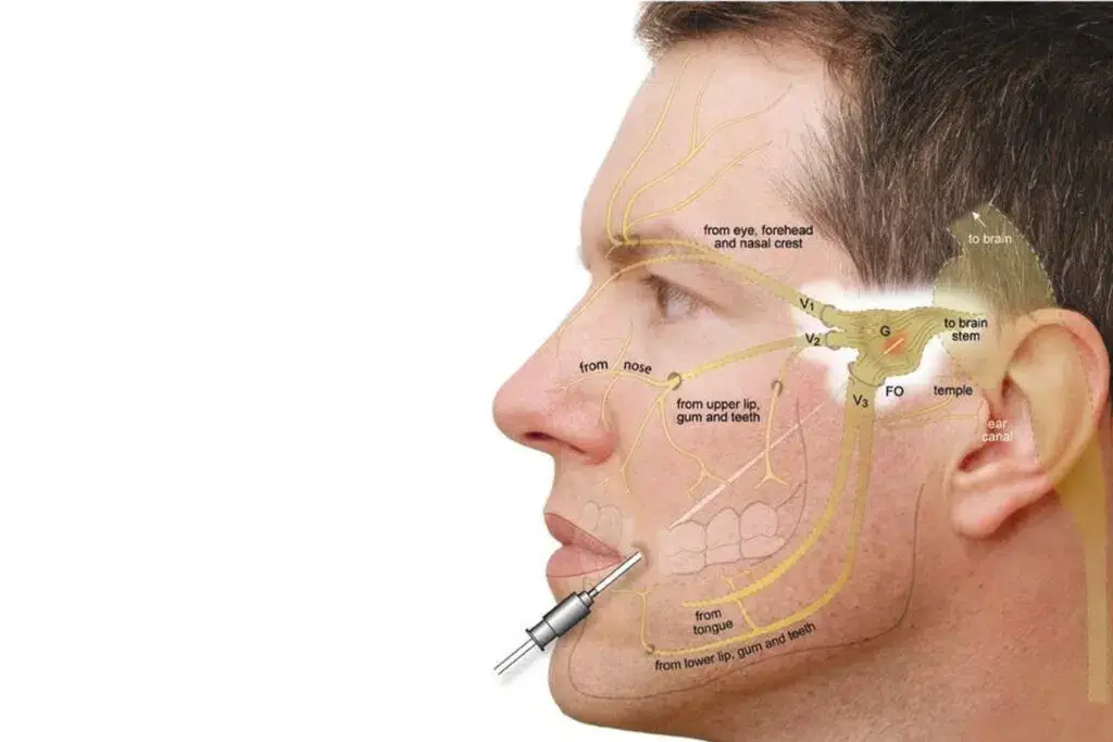
Pulsed RF is used anywhere a nerve must be treated but cannot be safely heated. The dorsal root ganglion (DRG) — the cell body cluster for spinal nerve roots — is a prime target. Heating the DRG risks permanent motor weakness; pulsed RF avoids this entirely. It is used for cervical and lumbar radiculopathy, occipital neuralgia (a frequently missed cause of the back-of-head pain that many patients in India chalk up to "tension"), trigeminal neuralgia, intercostal neuralgia, post-herpetic neuralgia, and complex regional pain syndrome (CRPS). It can also be applied near the shoulder, hip, knee, and sacroiliac joint as a gentler alternative when thermal ablation is inappropriate.
This is where PRF differs most from its thermal cousins. Onset can be slower — sometimes 4 to 8 weeks — because the mechanism is biological modulation rather than immediate nerve disruption. Duration is typically 6 to 12 months, though some patients report sustained benefit beyond 18 months, particularly for DRG applications. The procedure can be repeated without any accumulative nerve damage risk, which makes it uniquely suitable for younger patients.
Any modern RF generator capable of pulsed mode can deliver PRF. The key difference from thermal RFA lies in the generator settings and the physician's decision-making — not the hardware. Precision electrode placement, confirmed by sensory testing at appropriately low thresholds, is essential.
Where radiofrequency uses electrical current to generate heat, cryoablation turns physics on its head and uses cold — extreme cold — as its therapeutic tool. A specialised cryoprobe contains two concentric channels: pressurised gas (nitrous oxide or argon) flows through the inner channel to the probe tip, where it rapidly expands (the Joule-Thomson effect). This expansion drops the temperature at the probe tip to −70°C, freezing the surrounding tissue into a precise ice ball.
At this temperature, the water inside nerve cell bodies crystallises. Ice crystals rupture cellular membranes. The axons — the pain-conducting fibres — are destroyed. But crucially, the outer connective tissue sheath of the nerve (the epineurium and endoneurium) is preserved. This is the key biological distinction between cryoablation and thermal techniques. When a thermal RF procedure destroys a nerve, regrowth is somewhat chaotic. With cryoablation, the preserved sheath acts as a scaffold — axons regrow along their original path, predictably and cleanly. This is called Wallerian degeneration followed by guided regeneration, and it is the reason cryoablation is considered functionally reversible.
Cryoablation excels where reversibility and preservation of nerve architecture matter most. The genicular nerves of the knee — in patients with osteoarthritis who are awaiting or declining knee replacement — are an ideal target. There is good evidence for intercostal neuralgia, post-surgical chest pain (common after cardiac bypass or thoracic surgery), inguinal neuralgia (a cause of groin pain that is widely misdiagnosed), and facial neuralgias. It is increasingly used for peripheral nerve pain in the limbs.
One important clinical scenario: younger patients. For a 35-year-old IT professional in Hitec City with severe knee pain from a sports injury, I would strongly consider cryoablation over thermal RFA, precisely because I can offer long-lasting relief without committing to permanent nerve destruction in someone who will live with that decision for another forty years.
Relief typically begins within 1 to 2 weeks as post-procedure oedema settles. Duration of effect is 9 to 18 months, reflecting the time for axonal regeneration. The procedure can be repeated when pain returns. Interestingly, some evidence suggests that repeated freeze-thaw cycles create progressively more complete nerve disruption, potentially extending relief with subsequent treatments.
Cryoablation requires a dedicated cryotherapy system — the Iovera° device (Myoscience/Pacira) is a handheld system designed for peripheral nerve cryotherapy and has CE marking and significant clinical data behind it. The CryoPen and traditional large cryoprobes are used in different settings. The procedure typically requires ultrasound guidance for peripheral nerve targets, and fluoroscopy for spinal applications.
| Parameter | Thermal RFA | Cooled RFA | Pulsed RF | Cryoablation |
|---|---|---|---|---|
| Mechanism | Resistive heat, coagulation | Distributed heat via water cooling | EM field modulation, no heat | Freeze-induced axon destruction |
| Temperature | 80–90°C at tip | 60–80°C distributed | ≤42°C | −70°C |
| Lesion size | 5–7 mm cylinder | 8–12 mm sphere | No tissue lesion | Ice ball — variable, 10–20 mm |
| Onset of relief | 2–4 weeks | 2–6 weeks | 4–8 weeks | 1–2 weeks |
| Duration | 12–24 months | Up to 24 months | 6–12 months | 9–18 months |
| Reversible? | No | No | Yes | Yes (axons regrow) |
| Safe near motor nerve? | With caution | With caution | Yes | Peripheral only |
| Primary indications | Lumbar/cervical facets, knee | SI joint, hip, knee (COOLIEF) | DRG, occipital, radiculopathy | Knee, intercostal, inguinal, face |
| Guidance needed | Fluoroscopy | Fluoroscopy | Fluoroscopy / Ultrasound | Ultrasound / Fluoroscopy |
| Study / Trial | Modality | Indication | Key Finding |
|---|---|---|---|
| Dreyfuss et al., 2000 (Spine) | Thermal RFA | Lumbar facet pain | 60% of patients achieved ≥90% pain relief at 12 months |
| Patel et al., 2012 (PM&R) | Cooled RFA (INSITE) | Sacroiliac joint | 74% responders vs 17% sham at 3 months; durability to 24 months |
| Cahana et al., 2006 (Pain) | Pulsed RF | Chronic radiculopathy | Significant reduction in VAS at 3 months vs sham |
| Mikeladze et al., 2003 (Reg Anesth Pain Med) | Pulsed RF DRG | Lumbar radicular pain | 72% reported clinically meaningful improvement at 6 weeks |
| Trescot, 2003 (Pain Physician) | Cryoablation | Peripheral nerve pain | Comprehensive review — 80% success rate across multiple peripheral nerve targets |
| Davis et al., 2018 (Reg Anesth Pain Med) | Cooled RFA | Knee OA (COOLIEF) | 47% reduction in pain vs 24% with IA corticosteroid at 6 months |
| Falco et al., 2012 (Pain Physician) | Thermal RFA | Cervical facets | Level II evidence for cervical medial branch RFA; 8–24 months relief |
I want to say something directly — something I believe deeply after years of training in the UK and now eight years of practice in Hyderabad. Pain in India is undervalued. Dismissed. Medicalised with tablets and told to manage. Patients in their forties with severe facet joint disease are handed a muscle relaxant and told to do some exercises on YouTube. Teenagers with occipital neuralgia — real, debilitating pain at the back of the skull — are told it is exam stress and to "drink more water."
Yes, stress is a real contributor to pain. The cortisol released during repeated exam anxiety, the tension in neck muscles during twelve hours of NEET preparation, the postural collapse from sitting on a hard wooden chair with a stack of textbooks — all of this is physiology, not imagination. But the answer is not dismissal. The answer is understanding, accurate diagnosis, and appropriate intervention.
I see software engineers from HITEC City who sit in the same posture for ten hours, then sit in traffic on ORR for another two, then sit on a sofa watching OTT until 1 AM. Their cervical spine is enduring a daily punishment that no amount of "posture awareness" can fully undo. When that leads to cervical facet pain or occipital neuralgia, pulsed RF or thermal RFA of the appropriate target nerve can give them their life back — sometimes within a few weeks.
I see older patients — sixty-five, seventy — whose sacroiliac joints have been grinding away for years. They've been doing temple rounds for decades, long walks on uneven floors, sitting on the floor for religious ceremonies. Their SI joint pain is real, structural, and measurable on examination. Cooled RFA can dramatically reduce their pain and restore their independence.
The point is this: these procedures are not exotic Western interventions. They are evidence-based, minimally invasive tools that belong in every serious pain clinic in India. And more importantly, every patient deserves to know they exist.
The patient who benefits most from ablation procedures has:
A diagnostic medial branch block or joint injection — performed before any ablation — is mandatory in my practice. It is not a revenue-generating step. It is the only honest way to know whether the nerve we are planning to ablate is actually responsible for a patient's pain. I have seen patients elsewhere who had thermal RFA without prior diagnostic blocks and got no relief, because the wrong nerve was treated. This is avoidable.
Radiofrequency ablation uses a high-frequency alternating electrical current — delivered through a needle electrode — to generate heat at a precise tissue target. The heat disrupts pain-carrying nerve fibres, interrupting the pain signal before it reaches the brain. The procedure is done as a day-care intervention under fluoroscopy or ultrasound guidance, with no hospital admission required in most cases.
Thermal RFA typically provides 12 to 24 months of significant pain reduction. Cooled RFA for sacroiliac joint pain has demonstrated benefits up to 24 months in the published INSITE trial. Pulsed RF provides 6 to 12 months of neuromodulatory relief. Cryoablation lasts 9 to 18 months as nerve axons slowly regenerate. All can be safely repeated when the nerve recovers.
Yes, and this is non-negotiable in responsible practice. A diagnostic block — a small injection of local anaesthetic near the target nerve — is performed before any ablation. If the block produces significant, temporary pain relief (typically ≥50% or ≥80% depending on the guideline), it confirms that nerve is a meaningful pain source. Only then do we proceed to ablation. This step prevents unnecessary procedures and ensures the right target is treated.
Thermal RFA heats the tissue at the needle tip to approximately 80–90°C, producing a focused cylindrical lesion around 5–7 mm across. Cooled RFA circulates water internally through the probe tip, preventing immediate charring and allowing heat to distribute outward — creating a larger spherical lesion of 8–12 mm. This larger coverage is critical for targets like the sacroiliac joint, where nerve anatomy varies considerably between patients.
Yes — and this is the primary clinical reason to choose pulsed RF over thermal techniques near important nerve roots. Pulsed RF delivers energy in short bursts with long cooling intervals, keeping maximum tissue temperature below 42°C. This is below the threshold that damages nerve fibres structurally. It modulates pain signalling without causing motor weakness or permanent sensory loss — making it the correct choice for dorsal root ganglia, cervical radiculopathy, and occipital nerves.
Cryoablation freezes rather than heats. Pressurised gas expands at the probe tip, reaching −70°C. This creates a controlled ice ball that destroys pain-carrying axons through ice crystal formation — but crucially preserves the outer myelin sheath and connective tissue scaffolding of the nerve. Axons regenerate along this preserved sheath over 9–18 months — making the procedure functionally reversible. This distinguishes it clearly from thermal techniques, where structural nerve destruction is less predictably reversible.
Yes. IBAP Clinics — at Banjara Hills and Madeenaguda in Hyderabad — offers the complete spectrum of ablation procedures: thermal RF, cooled RFA, pulsed RF and cryoablation. All procedures are performed under specialist guidance by Dr. Vijay Bandikatla, who trained in the UK with an advanced pain fellowship from Cambridge University Hospitals and holds the FRCA and FFPMRCA from the Royal College of Anaesthetists, London. A diagnostic block always precedes any ablation procedure at our clinics.
Stop managing pain. Start treating it. Book a specialist consultation at IBAP Clinics — Hyderabad's dedicated interventional pain centre — and get a clear diagnosis and a personalised treatment plan.
This article has been written by Dr. Vijay Bhaskar Bandikatla (MBBS, DA, FRCA, FFPMRCA) for general educational purposes only. It does not constitute medical advice and should not be used as a substitute for professional clinical consultation. Suitability for any interventional procedure — including radiofrequency ablation, cooled RFA, pulsed RF, or cryoablation — must be determined by a qualified pain specialist following a comprehensive clinical assessment, diagnostic imaging review, and, where appropriate, diagnostic nerve block. Individual outcomes vary. IBAP Clinics, Hyderabad. © 2025 Vijay Advanced Pain Clinics Pvt. Ltd.
Banjara Hills
2nd Floor, 284/A, Road No. 12, above IDFC First Bank, near Omega hospitals, MLA Colony, Banjara Hills, Hyderabad, Telangana 500034.

Indo-British Advanced Pain Clinic © 2026. All Rights Reserved. Designed & Developed By Adroit
WhatsApp us