Helpline :
9807 55 6789
- Call Us Now: 9807 55 6789Call Us Now: 9807 55 6789
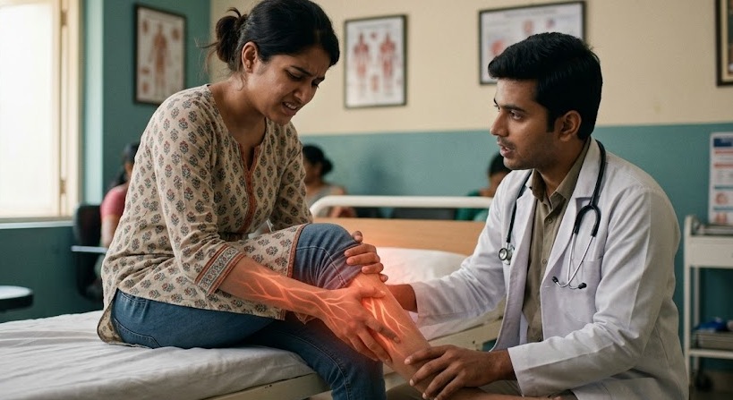
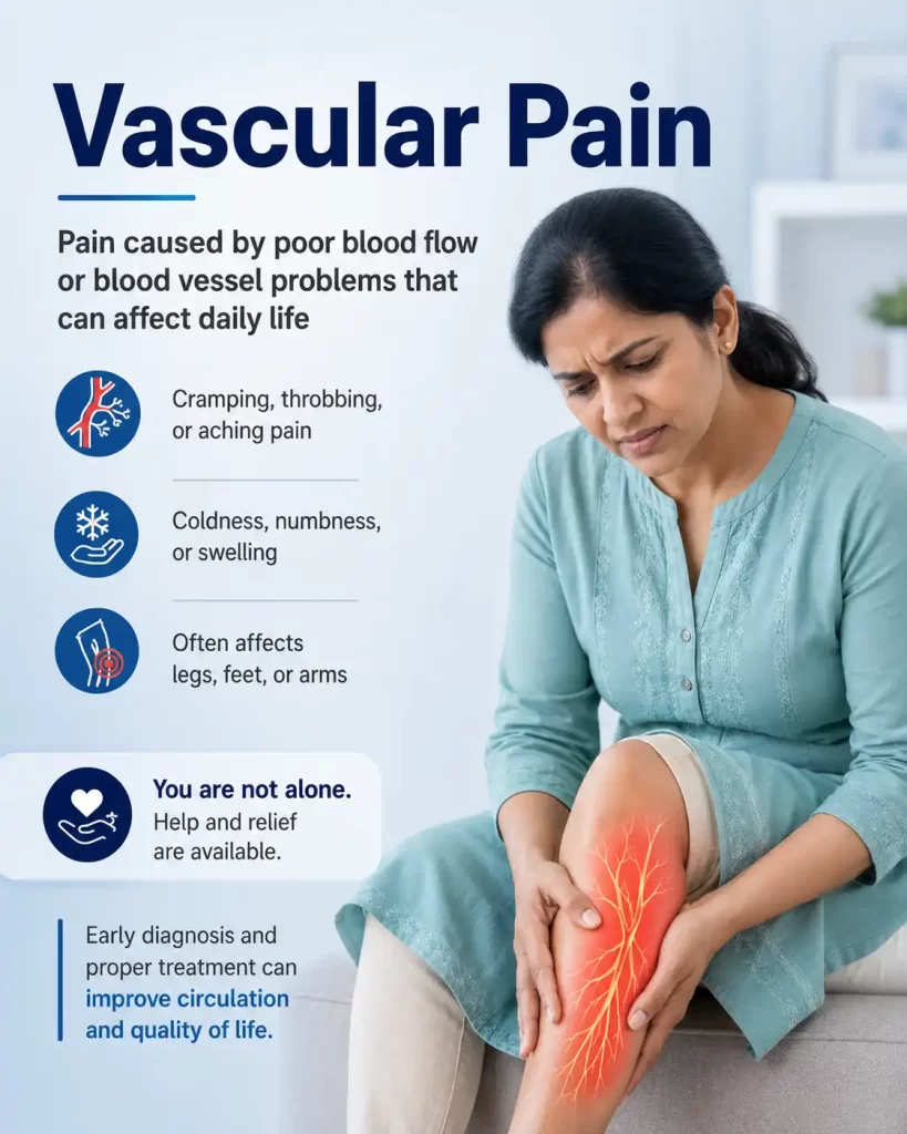
Vascular pain is not simply about blocked pipes. It spans a spectrum from the cold, dead-tissue ischaemia of arterial disease to the burning, over-perfused agony of erythromelalgia and understanding which direction the circulation has gone wrong determines everything about how we treat it. At IBAP Clinics, our mission is “Freedom from pain without surgery” delivering precision diagnosis and evidence-based, minimally invasive care for every point on this spectrum.
Think of your circulatory system as the road network of a city. Arteries are the highways high-pressure, outbound, carrying oxygen-rich blood to every organ and limb. Veins are the return roads, lower pressure, bringing deoxygenated blood back to the heart. When this network is functioning well, you do not feel it at all. Pain begins the moment the flow is either dangerously reduced or paradoxically, pathologically excessive.
In my clinic in Hyderabad, I see vascular pain presentations almost every week. A middle-aged software professional who has been smoking a pack a day for twenty years suddenly cannot walk more than 200 metres without his calves seizing up. A diabetic gentleman whose foot ulcer will not heal, not because the wound care is inadequate, but because there simply is not enough blood reaching the skin to carry the healing. A young woman who dreads summer because her feet turn scarlet and burn as if someone has put them in a hot furnace and the only relief is dunking them in a bucket of cold water.
These are all vascular pain problems. Very different in their mechanisms. Unified in one thing they are profoundly undertreated in most of India, because the connection between blood vessel health and chronic pain is still not well understood, even in many medical circles.
“Imagine Hyderabad’s Ring Road with two very different problems. Insufficient blood flow is like a road completely dug up no vehicles can reach their destination, and the neighbourhood suffers and withers. Excess vascular dilation is like all the traffic signals malfunctioning simultaneously too much chaotic blood floods the area, creating pressure, noise, heat, and congestion. Vascular pain medicine is, in essence, traffic management for your circulatory system.”
The pattern of symptoms is as important as the symptoms themselves. Vascular pain has a characteristic signature it is driven by ischaemia and inflammation in the vessel walls, activating nociceptors in a way that is quite distinct from musculoskeletal or purely neuropathic pain. Here is what to watch for.
| Symptom | Typical Features |
|---|---|
| Intermittent Claudication | Pain, cramping, or aching in the calves, thighs, or buttocks that starts with walking and subsides within minutes of rest — the most specific symptom of arterial disease |
| Burning or Throbbing | Sensations felt in the extremities, especially at night or when lying flat; also the hallmark of erythromelalgia |
| Skin Changes | Coolness, thin or shiny skin, hair loss, discolouration (pale, reddish-blue, or purple); paradoxically, vivid redness with warmth in erythromelalgia |
| Non-Healing Wounds | Sores on toes, feet, or ankles that do not close over weeks — a marker of critical limb ischaemia or severe venous hypertension |
| Heaviness / Dead Weight | Persistent feeling of heaviness in the affected limb disproportionate to activity; typical of chronic venous insufficiency |
| Pulse Deficits | Weak or absent pulses in the feet or wrists — a direct sign of arterial occlusion that every GP should check |
| Swelling, Redness, Warmth (acute) | Acute unilateral leg swelling with pain — must immediately raise suspicion of DVT, which is a medical emergency |
| Colour Changes in Fingers/Toes | White → blue → red sequence triggered by cold or stress — the classic triphasic response of Raynaud's phenomenon |
Sudden severe limb pain with a cold, pale, or blue extremity loss of pulse acute leg swelling with redness non-healing ulcers with black tissue or foul odour or any accompanying chest pain, breathlessness, or neurological deficits. These are vascular emergencies. Do not wait for a clinic appointment.
Vascular disorders are frequently progressive. The most common underlying culprit is atherosclerosis — the buildup of plaque (fat, cholesterol, and calcium) within arterial walls. But not all vascular pain is atherosclerotic. Here is the full landscape.
| Condition | What It Is | Typical Symptoms |
|---|---|---|
| Peripheral Artery Disease (PAD) | Narrowing of peripheral arteries, usually in the legs, restricting blood flow to muscles | Intermittent claudication, rest pain, non-healing ulcers |
| Deep Vein Thrombosis (DVT) | Blood clots in deep veins, often in the legs | Acute swelling, redness, warmth, pain; risk of pulmonary embolism |
| Chronic Venous Insufficiency (CVI) | Veins cannot return blood to the heart, leading to pooling and venous hypertension | Aching, heaviness, swelling, varicose veins, skin changes, ulcers |
| Raynaud's Phenomenon | Spasms in small arteries of fingers or toes, triggered by cold or stress | Colour changes (white → blue → red), numbness, burning |
| Buerger's Disease (TAO) | Inflammatory vasculitis of small/medium arteries, almost exclusively tobacco-driven | Ischaemia of digits, superficial thrombophlebitis, gangrene |
| Erythromelalgia | Pathological vasodilation — too much blood flooding the extremities | Burning pain, redness, warmth; relieved by cooling |
| Aneurysms | Abnormal bulges in blood vessels that may press on nerves or tissues, with rupture risk | Pulsating mass, deep aching pain; risk of acute rupture |
| Vasculitis | Systemic inflammation of blood vessel walls | Pain, tenderness, systemic features (fever, weight loss, fatigue) |
| Diabetic Ischaemic Disease | Accelerated macro- and microvascular disease in diabetes | Claudication, rest pain, non-healing foot ulcers, neuropathic overlap |
People worldwide affected by peripheral arterial disease
Of diabetics develop significant vascular complications
Higher limb amputation risk in smokers with PAD vs non-smokers
SCS success rate for inoperable critical limb ischaemia (pain relief)
Peripheral arterial disease (PAD) is, at its core, atherosclerosis of the limb arteries. Plaques of cholesterol, calcium, and fibrous tissue build up inside the vessel walls, narrowing the lumen the working space through which blood flows. The result is reduced oxygen delivery to the muscles and skin of the legs.
The classic presentation is intermittent claudication pain, cramping, or heaviness in the calf (or thigh, or buttock, depending on the level of disease) that begins predictably after a certain walking distance and resolves completely with rest within minutes. The analogy I use with patients is this: imagine your calf muscles as a factory running at partial power. At rest, that partial supply is enough. But the moment demand increases walking the factory runs short of fuel, acidosis builds up, and pain shuts the system down.
In more advanced stages, the pain arrives even at rest. Rest pain typically worse at night, when the heart is pumping less forcefully and gravity no longer assists flow signals that the arteries cannot deliver even baseline requirements. Tissue loss follows: ulcers on the toes or heel that refuse to heal, and in the worst cases, gangrene.
The Ankle-Brachial Index (ABI) is the single most important bedside test. ABI < 0.9 confirms PAD. ABI < 0.4 indicates critical limb ischaemia. An ABI > 1.3 in a diabetic patient (falsely elevated due to arterial calcification) should prompt toe pressure measurement instead.
Venous disease is the opposite problem in terms of pressure dynamics, but equally painful. Here, blood accumulates in the legs because the venous valves the one-way gates that prevent backflow become incompetent. Blood pools in the lower limbs, pressure builds within the veins, and the surrounding tissues become oedematous, inflamed, and eventually damaged.
The pain of chronic venous insufficiency (CVI) is characteristically dull and aching, worst after prolonged standing, and relieved by elevation. Unlike arterial pain, it worsens with heat which is why patients report legs feeling worse in the Hyderabad summer, when temperatures push above 40°C and the blood vessels dilate further.
Venous leg ulcers typically occur around the medial malleolus the inner ankle and are shallow, weeping wounds surrounded by hyperpigmented (brown-stained), indurated (hardened) skin. They are notoriously slow to heal and chronically painful. In India, many patients tolerate these for months, even years, before seeking specialist help, often relying on home remedies or local bandaging without addressing the underlying venous hypertension.
Buerger’s disease is different from atherosclerotic PAD in one critical way: it is not primarily a cholesterol problem. It is an inflammatory vasculitis the body’s own immune cells attacking and occluding small and medium-sized arteries and veins. And it is almost entirely driven by tobacco. Not just cigarette smoking bidis, chewing tobacco, and hookah are all established triggers. The pathology is insidious: segmental thrombosis within inflamed vessel walls, typically affecting the tibial, peroneal, radial, and ulnar arteries first.
The patients are younger than typical PAD patients often men in their thirties and forties who are heavy smokers. The presentation is dramatic: painful ischaemia of the fingers or toes, Raynaud’s-like episodes, superficial thrombophlebitis (inflamed, red cord-like veins under the skin), and rapidly progressive gangrene if smoking continues.
I want to say this plainly, because I see it in my practice and it genuinely distresses me: TAO is a self-inflicted tragedy that is almost entirely preventable and, if caught early enough, arrestable. The single most effective treatment is absolute, complete, permanent cessation of all tobacco use. I have seen patients who stopped smoking completely go on to heal, preserve their limbs, and live relatively normal lives. I have seen patients who could not stop lose fingers, then hands, then legs. There is no other disease in pain medicine where patient behaviour so directly determines outcome.
Erythromelalgia is, in some ways, the mirror image of PAD. Where PAD is too little blood reaching the tissue, erythromelalgia is too much an uncontrolled, pathological vasodilation of the small blood vessels in the extremities, flooding the skin with far more blood than the tissue needs or can manage. The result is intense burning pain, vivid redness, and warmth predominantly in the feet, though hands can be affected too.
The disorder can be primary (genetic often a gain-of-function mutation in the SCN9A gene encoding the Nav1.7 sodium channel, the same channel family involved in inherited channelopathies I deal with regularly in my neuromodulation practice) or secondary to underlying conditions including polycythaemia vera, essential thrombocythaemia, or as a drug-induced effect from calcium channel blockers and some chemotherapy agents.
Triggers are warmth, exercise, and dependency (lowering the limbs). Relief comes from cooling patients elevate their feet, stand on cold marble floors, or immerse them in cold water. In Hyderabad summers, these patients are particularly distressed. The management is genuinely challenging: aspirin at low doses can be effective in secondary erythromelalgia associated with thrombocytosis; sodium channel blockers (mexiletine, carbamazepine, lidocaine infusions) address the channelopathy; and in refractory primary cases, spinal cord stimulation has shown meaningful benefit.
Tobacco does at least three terrible things to blood vessels simultaneously. First, nicotine directly stimulates the sympathetic nervous system, causing immediate and prolonged vasospasm the vessels clamp down, reducing flow acutely. Second, the carbon monoxide in cigarette smoke displaces oxygen from haemoglobin, meaning the blood that does reach the tissue is carrying less oxygen than it should. Third, smoking accelerates atherosclerosis over years and decades the cholesterol plaques that narrow arteries develop faster, earlier, and more extensively in smokers.
The consequence in pain practice is that smokers present with ischaemic pain syndromes ten to fifteen years earlier than non-smokers, with much more aggressive disease progression, and with dramatically worse outcomes from both surgical and interventional treatments. Smoking doubles the rate of claudication progression to critical limb ischaemia. It obliterates the benefit of peripheral bypass surgery. In TAO, as I mentioned above, it is the disease itself.
Calf cramps deserve their own discussion because they cause enormous anxiety and are one of the most common reasons patients present to vascular and pain clinics asking whether they have “a blockage.” The majority do not.
Nocturnal calf cramps the classic scenario of being woken from sleep by a sudden, agonising muscle spasm in the calf are usually benign and not caused by vascular disease. They are far more commonly caused by muscle fatigue, dehydration, electrolyte imbalances (low magnesium, potassium, or calcium), prolonged sitting or standing, and lumbar nerve root compression. In pregnant women, they are extremely common and thought to relate to altered calcium and magnesium metabolism.
Claudication the arterial-ischaemic cramp is quite different in character. It is predictable (comes at a consistent walking distance), reproducible (the same every time), relieved by rest within two to five minutes (not by stretching or massage), and accompanied by other signs of arterial disease on examination. If you have cramps only at night, you probably do not have PAD. If you have cramps every time you walk 400 metres that vanish within three minutes of stopping, please see a doctor about your arteries.
Diabetes creates vascular pain through two simultaneous mechanisms: macrovascular disease (accelerated atherosclerosis of the large and medium arteries essentially an extreme form of PAD) and microvascular disease (damage to the tiny capillaries and arterioles that feed the skin and nerves). This is what makes the diabetic foot so uniquely challenging and dangerous.
The macrovascular disease causes the same claudication and rest pain as PAD. The microvascular disease adds a layer of neuropathic pain (from nerve ischaemia), impairs wound healing at a cellular level, and renders the skin extraordinarily vulnerable to infection. And because diabetic neuropathy can also blunt pain sensation, patients sometimes sustain injuries or develop ulcers without feeling them the pain system that would normally alert them to danger has been partially silenced.
Managing diabetic ischaemic pain requires exceptional HbA1c control, aggressive cardiovascular risk factor management, meticulous foot care education, and when conventional measures fail specialised pain interventions including lumbar sympathetic blocks and spinal cord stimulation.
This is where vascular pain medicine gets genuinely tricky. Leg and limb pain have many possible causes. A patient who cannot walk 300 metres might have claudication or spinal stenosis. The two feel superficially similar but respond to utterly different treatments. Getting this wrong wastes months and, frankly, exposes patients to interventions they do not need. At IBAP Clinics, we take the differential seriously here is how the key conditions separate out.
| Condition | Key Features | How It Differs from Vascular Pain |
|---|---|---|
Musculoskeletal Pain (strain, tendinopathy) | Worsens with specific movements, localised tenderness, persists at rest | Not relieved by rest alone; no skin changes; pulses normal |
Neuropathic Pain (radiculopathy, peripheral neuropathy) | Burning, tingling, numbness; often constant; follows nerve distribution | Not activity-dependent; associated with diabetes or spine disease; ABI normal |
Neurogenic Claudication (spinal stenosis) | Pain and weakness in legs with walking; relieved by sitting forward or bending | Relief with posture change, not just stopping; back pain prominent; pulses normal |
Exertional Compartment Syndrome | Tightness and pain during exercise; resolves with rest; mainly young athletes | No skin changes; pulses normal; does not follow walking-distance pattern |
Venous Insufficiency (vs arterial) | Aching, heaviness, swelling; worse at end of day; improves with elevation | Swelling and skin changes more prominent; ABI normal; no claudication pattern |
DVT (Deep Vein Thrombosis) | Acute unilateral swelling, pain, warmth, redness | Medical emergency; requires anticoagulation; risk of pulmonary embolism |
Referred Pain (hip/knee OA, sacroiliac joint) | Deep ache, limited range of motion, joint-specific signs | Imaging shows degenerative changes; pulses normal; no claudication pattern |
“The simplest screening question I ask every patient with leg pain is: does the pain come on at a predictable walking distance, and does it disappear completely within three minutes of stopping not just easing, but disappearing? If the answer to both is yes, I am already thinking arterial disease until proved otherwise. That single history point is worth more than any blood test.”
| Investigation | What It Tells Us | When We Use It | Limitation |
|---|---|---|---|
| Ankle-Brachial Index (ABI) | Ratio of ankle to arm systolic BP — screens for arterial disease | First-line for all suspected PAD | Falsely high in diabetics (calcified vessels) |
| Duplex Doppler Ultrasound | Arterial and venous flow, anatomy, valve function | PAD, CVI, DVT, varicose veins | Operator-dependent; limited for iliac/aortic |
| CT Angiography (CTA) | Detailed arterial map, stenosis localisation, planning | Pre-intervention planning for PAD/TAO | Radiation; contrast nephropathy risk |
| MR Angiography (MRA) | Arterial anatomy without radiation | Younger patients, renal impairment | Overestimates stenosis; slower |
| Treadmill Exercise Testing | Vascular response under physical stress — measures claudication distance | Unmasks borderline PAD; quantifies functional limitation objectively | Requires patient cooperation; unsafe in severe CLI |
| Photoplethysmography (PPG) | Blood circulation near the skin surface — detects small-vessel disease | Raynaud's phenomenon, erythromelalgia, digital ischaemia | Specialist equipment; limited availability in Hyderabad |
| Venous Duplex Ultrasound | Vein patency, reflux, valve competence | DVT diagnosis and chronic venous insufficiency staging | Operator-dependent |
| Toe Pressure / TcPO₂ | Distal perfusion; wound healing potential | Diabetic foot, critical limb ischaemia | Specialist equipment needed |
| Nerve Conduction Studies | Separates neuropathic from ischaemic pain component | Diabetic patients with mixed symptoms | Does not assess small-fibre neuropathy |
| Blood Tests (FBC, HbA1c, Lipids, Coag) | Risk factor profile, haematological causes | All patients; polycythaemia screen for erythromelalgia | Non-specific |
Optimal medical therapy for vascular pain involves targeting multiple pathways simultaneously. For arterial disease, antiplatelet therapy (aspirin, clopidogrel) reduces thrombotic risk, statins stabilise atherosclerotic plaques and have direct anti-inflammatory effects on vessel walls, and ACE inhibitors or ARBs improve endothelial function. Cilostazol a phosphodiesterase inhibitor has Level A evidence for improving claudication distance in PAD and is underused in India.
For venous disease, compression therapy remains the mainstay 20–40 mmHg graduated compression stockings worn daily. Venoactive drugs (diosmin, horse chestnut seed extract, oxerutins) have modest evidence for symptom control in CVI. Pentoxifylline improves microcirculation and has evidence for venous ulcer healing as an adjunct.
Supervised exercise therapy a structured walking programme that deliberately induces claudication pain at controlled intervals is one of the most evidence-based treatments for PAD and yet almost nobody recommends it in India. It works by developing collateral circulation, improving muscle oxygen extraction, and reducing cardiovascular risk.
Smoking cessation pharmacotherapy varenicline (Champix), NRT patches, or bupropion must be offered to every tobacco-using patient with vascular pain. Not as optional advice. As a formal prescription, written down, followed up at every visit. The pain simply will not respond adequately to anything else we do while smoking continues.
For patients with arterial blockages, restoring vessel patency is the priority. These procedures are performed under local anaesthesia with sedation, often as day-care, guided by fluoroscopy or ultrasound they are not open surgery.
| Procedure | Indications | Benefits | Key Risks |
|---|---|---|---|
| Angioplasty | PAD with focal stenosis or short-segment occlusion | Opens narrowed arteries, improves blood flow, rapid recovery | Bleeding, vessel dissection, re-narrowing (restenosis) |
| Stenting | Recurrent stenosis after angioplasty; long lesions | Keeps arteries open for sustained flow restoration | In-stent restenosis, thrombosis, rare migration |
| Thrombolysis / Thrombectomy | Acute DVT or arterial thrombosis; limb-threatening ischaemia | Dissolves or removes clot; limb salvage | Bleeding, distal embolisation |
When vascular pain is driven by sympathetic overactivity, central sensitisation, or persists despite restored flow, the interventional pain toolkit becomes essential. These are not last resorts they are specialist tools that, when applied at the right stage, can dramatically change outcomes.
| Procedure | Target | Best For | Evidence Level | IBAP Expertise |
|---|---|---|---|---|
| Lumbar Sympathetic Block | L2–L4 sympathetic chain | PAD, TAO, ischaemic rest pain, diabetic foot, CRPS lower limb | Strong | ✓ Specialist |
| Lumbar Sympathetic Ablation (RFA) | L2–L4 ganglia (thermal) | Longer-term effect where diagnostic blocks are effective | Good | ✓ Specialist |
| Stellate Ganglion Block | Cervico-thoracic sympathetic | Upper limb TAO, Raynaud's, erythromelalgia (arm), CRPS upper limb | Moderate | ✓ Specialist |
| D1/D2 Thoracic Sympathetic Ablation | T1–T2 ganglia | Upper limb hyperhidrosis, Raynaud's, TAO (hands) | Moderate | ✓ Specialist |
| Pulse RFA (Pulsed Radiofrequency) | Peripheral nerves or sympathetic chain | Neuralgia, ischaemic neuropathic pain; CRPS with sensitisation | Emerging | ✓ Specialist |
| Thermal RFA | Specific sympathetic ganglia | Refractory vascular pain; pain persisting despite improved flow | Good | ✓ Specialist |
| Epidural Infusion / Catheter | Epidural space (LA ± opioid) | Acute ischaemic crisis, ulcer debridement, TAO flares | Good | ✓ Specialist |
| Selective Nerve Root Block | Specific lumbar nerve roots | Radicular pain component in mixed vascular-neuropathic presentations | Good | ✓ Specialist |
| Spinal Cord Stimulation (SCS) | Dorsal columns T8–T10 | Critical limb ischaemia, inoperable PAD, TAO, refractory CRPS | Strong | ✓ Specialist |
| Intrathecal Drug Delivery (Pump) | Spinal fluid — direct drug delivery | Severe intractable CLI pain; failed SCS; contraindication to SCS | Good | ✓ Specialist |
The lumbar sympathetic chain runs along the anterolateral surface of the lumbar vertebral bodies (L1–L4), and it carries both pain signals from the lower limb vasculature and efferent signals that maintain arterial tone and cause vasospasm. Interrupting this chain with a targeted injection of local anaesthetic under fluoroscopic or ultrasound guidance does two powerful things simultaneously: it reduces ischaemic pain by blocking afferent nociceptive signals, and it causes vasodilation of the leg arteries by removing the sympathetic vasoconstrictor drive, improving blood flow.
Typical side effects are mild and transient temporary leg heaviness or a slight drop in blood pressure. Serious complications (nerve injury, intravascular injection) are rare when performed by an experienced specialist using image guidance. The procedure is outpatient.
The stellate ganglion block is its upper-limb equivalent targeting the cervicothoracic sympathetic ganglion to achieve vasodilation in the arm and hand. It is the primary intervention for Raynaud’s phenomenon, upper-limb TAO, and CRPS of the hand. Temporary hoarseness and a droopy eyelid (Horner’s syndrome) are expected transient effects, not complications. Where diagnostic blocks show sustained benefit, RFA of the lumbar or thoracic sympathetic ganglia provides longer-lasting relief measured in months rather than days.
Radiofrequency ablation uses heat generated from radiofrequency waves to interrupt pain signals from specific nerves or ganglia. It is particularly valuable when pain persists despite improved blood flow a situation that indicates central or peripheral sensitisation, or a CRPS-like overlay, rather than simple ongoing ischaemia. RF ablation delivers 6–18 months of relief in most responders and can be repeated. It is minimally invasive, outpatient, and reduces medication dependence significantly.
SCS occupies a unique position in vascular pain management it is one of the few interventions with Level A evidence specifically for critical limb ischaemia in patients who cannot undergo surgical revascularisation. A systematic review and the Cochrane analysis both demonstrate significant improvements in rest pain, transcutaneous oxygen tension, ulcer healing, and limb salvage rates compared to conservative management alone.
Modern systems including high-frequency SCS (10 kHz) and dorsal root ganglion (DRG) stimulation deliver paresthesia-free relief with better targeting for focal limb pain. A trial period of 5–7 days before permanent implantation ensures that only responders proceed, which keeps the success rate high and patient satisfaction genuinely impressive.
“The spinal cord stimulator is a dimmer switch for the pain signals coming from your leg. It does not cure the arterial disease but it turns down the volume of pain significantly. And as a bonus, it seems to tell the blood vessels to relax and open up a little, which is why some patients actually see their ulcers begin healing after implantation. It is not magic, but for the right patient, it can genuinely transform their quality of life.”
For severe, intractable vascular pain where all other interventions have failed, an intrathecal pump delivers medication (morphine, ziconotide, or local anaesthetic) directly into the spinal fluid achieving high-efficacy pain relief with a fraction of the systemic drug dose. This means far fewer side effects than oral opioids. The pump is programmable and titratable, and the indications are specific: refractory rest pain in critical limb ischaemia, or severe CRPS where SCS has failed or is contraindicated. The goal is to avoid amputation, avoid high-dose systemic opioids, and preserve quality of life.
Regenerative therapies aim to support tissue repair, reduce localised inflammation, and in some contexts improve microvascular circulation. They are most valuable as adjuncts to the primary vascular and pain intervention, particularly for chronic wounds and ischaemic tissue.
| Therapy | Best Indications | Benefits | Key Considerations |
|---|---|---|---|
| Platelet-Rich Plasma (PRP) | Non-healing ulcers, ischaemic tissue inflammation, CRPS | Natural growth factors, anti-inflammatory, may accelerate wound healing | Pain at injection site; requires good evidence base for the specific application |
| BMAC / Mesenchymal Stem Cells | Severe tissue loss, refractory ulcers, promoting angiogenesis | Mesenchymal stem cells may promote new vessel formation; autologous, safe | More invasive harvest; variable evidence; cost |
| Nanofat / Microfat Injection | Skin quality improvement, microcirculation support in ischaemic zones | Minimally invasive, autologous, cosmetic and functional benefit | Swelling, bruising; rare nodules |
Start your journey with a virtual consultation to discuss symptoms from home.
We review your medical history and relevant reports for a clear understanding.
Our doctors conduct a thorough assessment through detailed discussions.
We confirm findings with state-of-the-art imaging like X-rays, CT scans, and MRIs.
Our team identifies the root cause and key trigger points for treatment.
We create a customized treatment plan, including necessary medications and procedures.
Our Pain Specialists support a complete recovery focused on total wellness.
We provide ongoing follow-ups tailored to each treatment plan, ensuring continuous care and long-term recovery support.
We relieve your pain, helping you be yourself again!
Arterial pain arises from inadequate blood supply it cramps during walking and resolves completely with rest. Venous pain comes from blood pooling and pressure in the veins it is a dull, aching heaviness that worsens with prolonged standing and improves when you elevate your legs. The distinction is clinically critical because the treatments are entirely different.
Absolutely and this is one of the most underappreciated facts in vascular medicine. In Buerger’s disease (TAO), smoking is not just a risk factor it is the direct cause of the inflammatory vasculitis that destroys the blood vessels. TAO typically affects men and women in their thirties and forties. Stopping smoking completely is the only way to halt the disease.
A lumbar sympathetic block is a precise injection around the sympathetic nerve chain in the lumbar spine, performed under X-ray guidance. It simultaneously blocks pain signals from the ischaemic tissue and causes blood vessel dilation by removing the sympathetic vasoconstrictor drive improving both pain and circulation in a single procedure. It is the cornerstone interventional treatment for PAD, TAO, and diabetic ischaemic rest pain.
It is possible. Erythromelalgia causes intense burning pain with visible redness and warmth the opposite of poor circulation. It worsens with heat and exercise, and improves with cooling. It is frequently misdiagnosed for years. If cooling relieves your symptoms and warmth reliably triggers them, please seek specialist evaluation. Blood tests including a full blood count are important to rule out polycythaemia vera as an underlying cause.
Usually not. Nocturnal calf cramps are most often benign caused by dehydration, magnesium deficiency, prolonged sitting, or nerve root irritation in the lumbar spine. Arterial claudication (ischaemic calf cramps) occurs during walking at a consistent distance, not at night, and resolves fully within minutes of stopping. If your cramps are exclusively nocturnal and have no relationship to walking, vascular disease is less likely but a simple ABI test will reassure you definitively.
SCS modulates sympathetic outflow to the peripheral blood vessels (similar to a repeated sympathetic block) and directly suppresses ischaemic pain signals in the dorsal horn. For patients with critical limb ischaemia who cannot undergo bypass surgery or angioplasty, SCS has Level A evidence for reducing rest pain, improving skin perfusion, and in some cases aiding ulcer healing and preventing amputation.
Start with an Ankle-Brachial Index (ABI) a quick, non-invasive bedside test. A Duplex Doppler ultrasound assesses both arterial and venous flow. CT or MR angiography provides detailed arterial mapping for planning interventions. Blood tests including HbA1c, lipid profile, and full blood count are routine. If neuropathic pain is suspected alongside the vascular component, nerve conduction studies help clarify the picture.

EXCELLENTTrustindex verifies that the original source of the review is Google. Good pain relief with treatmentPosted on GoogleTrustindex verifies that the original source of the review is Google. Best for pain reliefPosted on GoogleTrustindex verifies that the original source of the review is Google. बहुत अच्छा लगा हमें ये ट्रिटमेंट करा के अच्छे डॉक्टर और अच्छे स्टाफ हैं और काम पैसे में अच्छा अच्छा ट्रिटमेंट होता हैPosted on GoogleTrustindex verifies that the original source of the review is Google. Very nice, good and helpful staf, overall good experiencePosted on GoogleTrustindex verifies that the original source of the review is Google. Very good doctors and good tritment Thank you doctorPosted on GoogleTrustindex verifies that the original source of the review is Google. One of the best clinic for the relief of back painPosted on GoogleTrustindex verifies that the original source of the review is Google. Very good treatment indo british clinic for every pain reliefPosted on GoogleTrustindex verifies that the original source of the review is Google. I have visited indo british pain clinic this is superb clinic for pain relief that's why I have given 5 start rating to this clinic ...Posted on GoogleTrustindex verifies that the original source of the review is Google. My name is Raja Sekhar Reddy Tummuri from kakinada city My father was suffering nervous pain from last 3 years … I have decided to join with this op … Now everything fine after treatment done from DR. vijay sir … Wonderful service received from INDO British advanced pain clinic Hospitals in Hyderabad @ Banjara Hills road no 12.. Each and every staff response , concern , support & everything it’s too good Thank you so much … Special thanks to Mr. DR. Vijay bhaskar Bandi Katla …👏🏻👏🏻👏🏻👏🏻🙏🙏Verified by TrustindexTrustindex verified badge is the Universal Symbol of Trust. Only the greatest companies can get the verified badge who has a review score above 4.5, based on customer reviews over the past 12 months. Read more
Medical Disclaimer: This article is written for educational purposes by Dr. Vijay Bhaskar Bandikatla, Interventional Pain Specialist, IBAP Clinics Hyderabad. It does not constitute medical advice and should not replace a formal clinical consultation. Vascular pain conditions require individualised assessment, investigation, and treatment planning by a qualified clinician. If you are experiencing symptoms described in this article, please seek prompt medical evaluation. All interventional procedures mentioned carry risks that must be discussed with your treating physician in the context of your specific medical history.

MBBS, DA, FRCA (UK), FFPMRCA (Pain Medicine, RCOA, UK)
CCT (Anesthesiology And Pain Management)
Neuromodulation & Advanced Pain Research Fellowship (London), MBA (HM)
Banjara Hills
2nd Floor, 284/A, Road No. 12, above IDFC First Bank, near Omega hospitals, MLA Colony, Banjara Hills, Hyderabad, Telangana 500034.

Indo-British Advanced Pain Clinic © 2026. All Rights Reserved. Designed & Developed By Adroit
WhatsApp us