Helpline :
9807 55 6789
-
Call Us Now: 9807 55 6789Call Us Now: 9807 55 6789
A patient guide to plantar fasciitis: why it happens, why it persists, and how we treat it precisely at IBAP Clinics, Hyderabad.
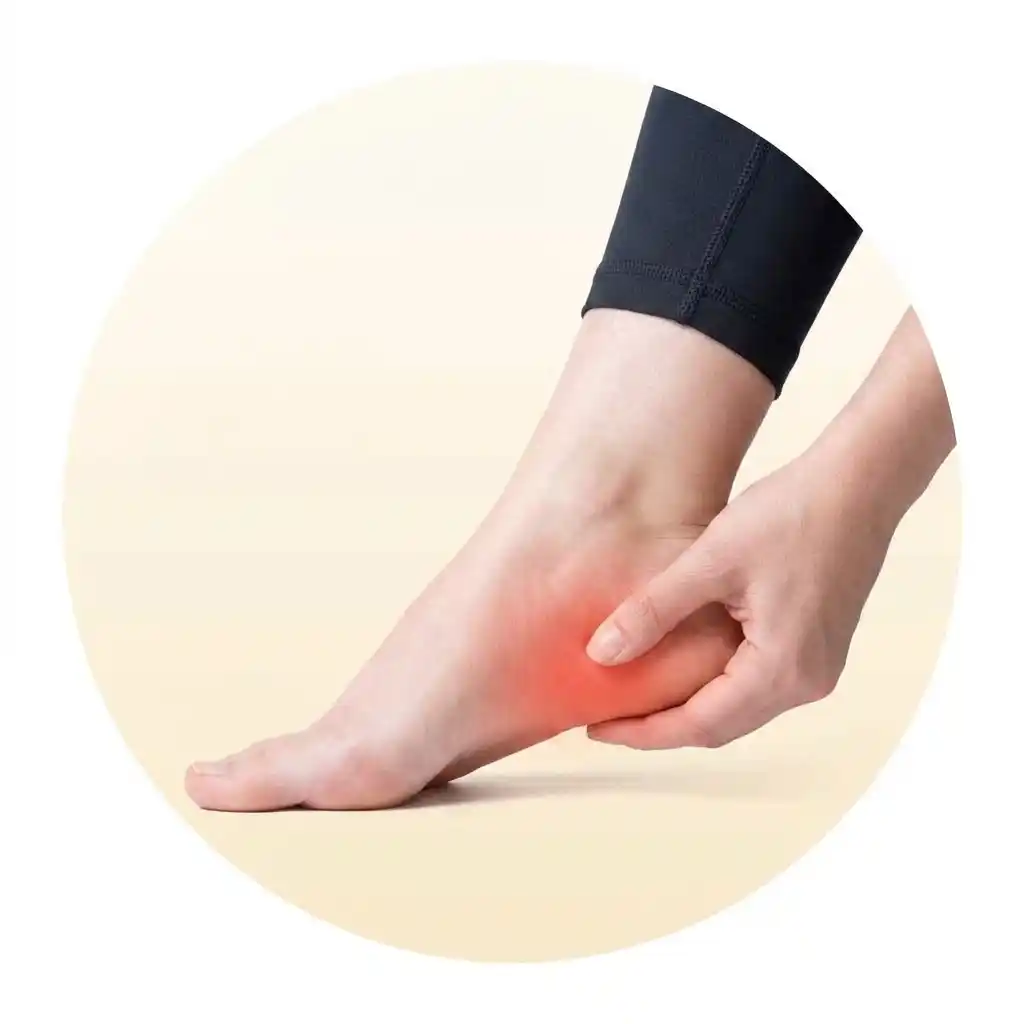
Non-surgical and effective care for lasting relief.
FIX AN APPOINTMENT
You wake up. The morning is quiet. And then your foot touches the floor, and there it is — a sharp, almost electric pain in the heel. It catches you off-guard every single time, even though you’ve had it for months. You limp to the bathroom. After ten minutes of walking it begins to ease. You think, maybe today will be better. Then you sit for an hour, get up again, and the same pain greets you.
This is plantar fasciitis. And it is one of the most common, most frustrating, and — I say this as a pain specialist — most frequently dismissed foot conditions I see in my clinic. Patients come to me after months of being told to just rest, or to “lose some weight,” or that it will sort itself out. Sometimes it does. But very often it doesn’t, and by the time they find their way to us, a straightforward condition has become a chronic, deeply entrenched pain pattern that now requires more intensive care to reverse.
I want to use this article to explain, clearly and honestly, what plantar fasciitis is, why it happens so commonly in Indian life specifically, what the real range of treatments looks like — from simple stretching right through to regenerative injections — and why early, accurate treatment is almost always better than waiting and hoping.
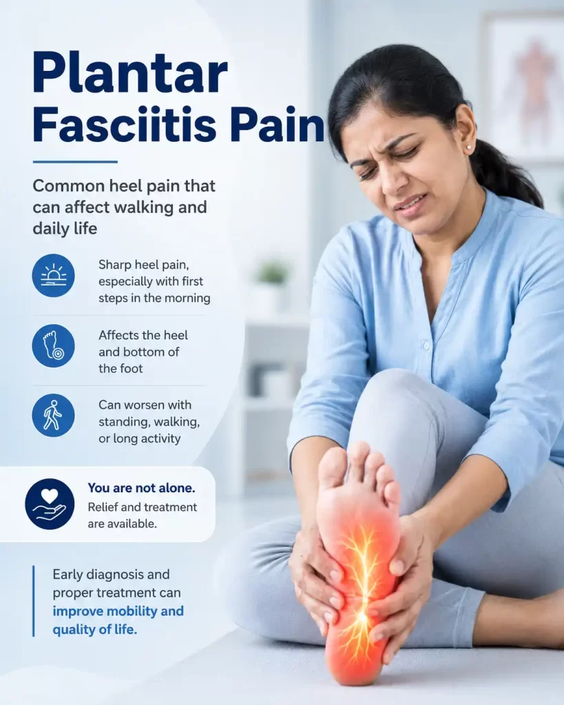
The plantar fascia is not a passive structure. It is a dynamic, load-bearing band under constant tension. When it begins to degenerate rather than just inflame, it stops responding to simple rest — and requires active biological stimulation to heal. This is why many cases that ignore early treatment become genuinely difficult to resolve later.
Think of the plantar fascia as a thick rubber band that runs along the underside of your foot, from the heel bone (calcaneus) all the way to the base of your toes. With every step you take, this band tightens slightly, stores elastic energy, and then releases it as you push off. It acts as a biological spring — shock absorber and energy recycler, both at once.
Now imagine that rubber band being stretched and contracted thousands of times a day, day after day, especially when the load is heavier than it was designed for, or when the mechanics of the foot are not quite right. Over time, tiny micro-tears accumulate at the point where the fascia anchors onto the heel. The body attempts to repair these tears — but if the load continues without sufficient recovery, the repair process falls behind. The tissue begins to degenerate rather than simply inflame. It thickens. It becomes disorganised at a cellular level. And it hurts — particularly after periods of rest, when it has contracted and then is suddenly asked to stretch again.
Imagine a worn-out bridge suspension cable that has had tiny wire breaks accumulating over years of heavy traffic. The cable is still holding — just. But every morning when the first heavy lorry rolls over it, you hear the creak and feel the sway. That is your plantar fascia. The problem isn’t just the weight of the lorry today — it’s the cumulative micro-damage that has built up while nobody was paying attention. Treating plantar fasciitis properly means reinforcing that cable, not just reducing the lorry’s speed.
I am asked this question at nearly every consultation for heel pain. It deserves an honest answer. A calcaneal spur — the bony outgrowth visible on an X-ray at the heel’s undersurface — is present in a large proportion of patients with plantar fasciitis. It looks dramatic on imaging. And naturally, patients assume it must be what is causing the pain.
But here is the thing: calcaneal spurs are equally common in people with no heel pain whatsoever. They form as a result of chronic tension and traction at the fascial insertion — the body’s attempt to stabilise a repeatedly stressed attachment site by laying down bone. The spur is a marker of long-standing mechanical stress. It is largely a bystander. Removing it surgically has not been shown to reliably resolve plantar fasciitis, which is why we do not do so. We treat the inflamed, degenerating fascia — and the underlying mechanical reasons it became inflamed in the first place.
People will experience plantar fasciitis in their lifetime
Patients improve with conservative care — if caught and treated early
Weeks for maximal benefit after shockwave or PRP treatment
Need surgery when regenerative options are used appropriately
I want to spend some time here, because I think this is important and is not covered enough in standard medical content. The Indian context — the specific pressures, habits, and environments of our daily lives — creates a near-perfect storm for plantar fasciitis.
Consider the commute. Hyderabad’s traffic is notorious. Many of our patients spend an hour each way in stop-and-go traffic, clutch-heavy driving, or crammed into shared autos and TSRTC buses. Then they spend eight to ten hours on hard floors — whether that is a nurse on a hospital ward, a software engineer who barely moves from their chair but then suddenly is on their feet all day during a site visit, a teacher standing on hard school floors, or a homemaker whose entire day is spent on marble or granite tiles that offer zero cushioning. The hard, flat surfaces that are standard in Indian construction — beautiful, cool, practical — are genuinely unkind to feet.
And then there is footwear. Traditional Indian footwear — chappals, kolhapuris, flat sandals — is culturally beloved and deeply practical for heat. But it provides almost no arch support and very little shock absorption. Wearing flat footwear on hard floors for hours at a time is one of the most reliable ways to overload the plantar fascia over time.
I also see a significant number of patients who have recently started walking programmes — on doctor’s advice for blood sugar or weight control — in footwear that is simply not designed for sustained walking. They go from near-zero daily steps to 5,000 or 8,000 steps a day, wearing the chappals they have always worn. The plantar fascia, unprepared and unsupported, takes the full brunt of this sudden increase in load.
Weight is another factor we must address honestly. Obesity and overweight are significant risk factors for plantar fasciitis — not because of moral failing, but because every kilogram of excess weight multiplies the load on the plantar fascia with every single step. India’s rising rates of metabolic syndrome, diabetes, and obesity are directly reflected in the caseload we see in clinic. And diabetes, beyond the weight factor, also changes the quality of connective tissue itself — making the fascia more prone to degeneration and slower to heal.
There are other, subtler contributors too. I see patients whose daily temple visits — where footwear is removed and they stand barefoot on hot, hard stone floors for extended periods — have contributed significantly to their heel pain. I see IT sector employees who sit in essentially the same posture for ten hours, then suddenly play a strenuous cricket match on the weekend and wonder why their heel is agony by Monday. Poor sleeping postures and prolonged sitting with feet in awkward positions can tighten the calf and Achilles complex, increasing fascial tension. Even the ubiquitous speed breaker — aggressive, inconsistent, sometimes unmarked — creates sudden, uneven loading through the foot every time it is cleared at anything above a crawl.
One thing I believe deeply is that validating a patient’s pain is not just a nicety — it is the beginning of effective treatment. When someone has been limping for eight months and their GP said “it’s just a heel spur, it will go away,” the psychological burden of unacknowledged pain is real and measurable. Chronic pain that is not taken seriously does not tend to resolve. It tends to entrench. Taking the time to say “yes, this is real, here is exactly what is happening, and here is what we are going to do about it” — that itself is therapeutic.
Some of the clearest risk profiles I see include people with excess body weight or obesity; those in jobs requiring prolonged standing or hard-surface walking — healthcare workers, teachers, retail staff, factory workers; middle-distance runners and those who have abruptly increased physical activity; people with structural foot issues such as flat arches (pes planus) or very high arches (pes cavus); those with tight calf muscles and reduced ankle flexibility; patients with diabetes or other metabolic conditions; and those who have been wearing poorly supportive footwear for years. But I want to be clear — plantar fasciitis does not discriminate neatly. I see it in slim, young software professionals who spend all day in a chair and all weekend playing sport. I see it in older patients whose heels have simply accumulated decades of load. I see it in postpartum women whose weight and biomechanics changed rapidly. The pattern matters less than the fact that the fascia is struggling — and needs help.
A diagnosis of plantar fasciitis is largely clinical — the history and pattern are often quite telling. But I will be honest with you: clinical impression alone is not sufficient justification for an invasive procedure. Before we inject anything into your heel — whether it is a corticosteroid, PRP, or any other agent — I want to see exactly what we are dealing with.
High-resolution musculoskeletal ultrasound is our standard diagnostic tool for heel pain at IBAP Clinics. Ultrasound allows us to visualise the plantar fascia directly, measure its thickness at the insertion point (healthy fascia is typically below 4 mm; inflamed, degenerating fascia is often 5–8 mm or more), identify focal tears or areas of degeneration, assess blood flow within the tissue (hypervascularisation is a key marker of active chronic degeneration), and importantly, rule out other diagnoses that can mimic plantar fasciitis — heel fat pad syndrome, tarsal tunnel syndrome, accessory muscles, bursitis, stress fracture of the calcaneus.
This distinction matters enormously. A tarsal tunnel syndrome will not respond to plantar fascia injections. A fat pad atrophy problem requires a different approach altogether. Getting the diagnosis right before committing to treatment is basic good medicine — and it is not how heel pain is always managed, even in well-resourced settings.
I want to walk through these in order, because I think there is often a false binary presented to patients: either you try rest and some home exercises, or you need a jab. The reality is a carefully tiered continuum of escalating options, and we use each layer intelligently based on how the condition is responding.
The very first thing we do is explain. Not just tell — explain. When a patient understands why their heel hurts, what is happening in the tissue, and how their activities are either loading or unloading the fascia, they become active participants in their own recovery rather than passive recipients of treatment. This is evidence-based — patient education has measurable impact on outcomes for musculoskeletal pain conditions. We advise temporary reduction in high-impact activities (not complete rest, which can cause its own problems), a graded return to walking and exercise, and specific attention to surface and footwear.
Supportive footwear with a cushioned heel, firm arch support, and a slight heel raise is often the single most impactful change a patient can make in the first week of treatment. For patients with significant flat feet or high arches, custom or prefabricated foot orthoses help redistribute load away from the fascial insertion. Night splints — which hold the ankle and foot in a slightly dorsiflexed position during sleep — have good evidence for reducing that characteristic morning pain by preventing the fascia from contracting overnight.
This is genuinely one of the most powerful tools available, and it is free. The plantar fascia-specific stretch — pulling the toes back towards the shin before taking any weight on the foot — has robust randomised trial evidence supporting its use. Calf stretching (both with the knee straight and slightly bent to target both the gastrocnemius and soleus muscles) reduces the tension transmitted to the fascial insertion through the Achilles chain. Progressive intrinsic foot muscle strengthening, eccentric loading protocols, and proprioceptive exercises help restore the mechanical environment the fascia needs to heal.
Soft-tissue mobilisation of the calf, deep tissue massage of the plantar fascia itself, and instrument-assisted soft tissue work can meaningfully reduce local stiffness and fascial tension. These work best as an adjunct to active rehabilitation rather than as standalone treatments.
Not to be confused with diagnostic ultrasound — therapeutic ultrasound delivers mechanical vibration energy at a frequency that penetrates soft tissue, generating gentle warmth, increased local circulation, and modulation of the inflammatory response. It is most useful in the early to subacute phases of the condition, as an adjunct to exercise.
IFT uses medium-frequency alternating currents delivered through the skin to reduce pain signalling and muscle guarding in the surrounding structures. It is a useful adjunct for pain modulation during the rehabilitation phase, particularly in patients who find direct loading exercises difficult in the early stages.
LLLT uses specific wavelengths of light to stimulate cellular metabolic processes, reduce inflammation at a molecular level, and promote local tissue repair. There is growing evidence for its use in plantar fasciitis as part of a multimodal programme — it is particularly useful in patients who need pain reduction to be able to engage with rehabilitation exercises effectively.
This is one of my preferred non-invasive options for plantar fasciitis that has been present for more than six weeks and has not adequately responded to the above. Extracorporeal shockwave therapy delivers high-energy acoustic waves into the tissue, stimulating neovascularisation (new blood vessel formation), disrupting pain signalling pathways, and triggering a controlled healing response in degenerative tissue. Radial shockwave disperses energy across a broader zone and is particularly useful for superficial, diffuse fascial involvement. Focused shockwave penetrates more deeply and targets more discrete lesions — useful when the degeneration is highly focal at the insertion. Several sessions are typically needed, and maximal benefit develops over six to twelve weeks as the tissue responds.
| Treatment | Evidence Level |
Best Timing | Key Advantage | Limitations |
|---|---|---|---|---|
| Stretching + exercise programme |
Strong (Level I) |
First-line, all stages |
No cost, high long-term durability |
Requires consistency and patient adherence |
| Orthotics & footwear |
Strong | First-line, chronic |
Biomechanical root- cause correction |
Custom devices carry cost; compliance variable |
| Manual therapy / massage |
Moderate | Adjunct, early– subacute |
Immediate symptom relief, improved mobility |
Best as adjunct, not standalone |
| Therapeutic ultrasound (physio) |
Moderate | Adjunct, early phase |
Non-invasive, safe, promotes healing |
Limited standalone evidence |
| IFT (Interferential Therapy) |
Moderate | Adjunct, pain phase |
Pain modulation, enables rehab participation |
Adjunctive only |
| Low-level laser (LLLT) |
Moderate– Good |
Adjunct, any stage |
Anti-inflammatory, supports tissue repair |
Requires multiple sessions |
| Shockwave therapy (ESWT) |
Strong (Level I) |
Subacute to chronic (>6 wk) |
Stimulates healing; non-invasive; durable |
3–5 sessions needed; response over 6–12 weeks |
| USG-guided PRP | Good (Level II) |
Chronic, refractory |
Biological regeneration using own growth factors |
Response time 6–12 weeks; not NHS- covered |
| Prolotherapy | Moderate | Chronic, ligamentous laxity |
Strengthens fascial attachment |
Multiple sessions; variable evidence base |
| Percutaneous tenotomy / fenestration |
Good | Chronic degeneration |
Resets degenerative tissue; less invasive than surgery |
Requires skilled USG operator; post- procedure rehab |
| USG-guided corticosteroid |
Good (short-term) |
Severe acute flare |
Rapid pain reduction | Risk of fascia rupture if overused; not regenerative |
| Feature | Radial Shockwave (rESWT) |
Focused Shockwave (fESWT) |
|---|---|---|
| Energy distribution | Broad, dispersed zone | Precise, deep focal point |
| Penetration depth | Superficial–intermediate (~3 cm) |
Deep (>3 cm) |
| Best for plantar fasciitis | Diffuse insertion tenderness, superficial pathology |
Focal degeneration at deep heel insertion |
| Pain during treatment | Moderate; well-tolerated |
Slightly more intense; usually manageable |
| Sessions required | 3–5 typically | 3–5 typically |
| Device availability | Widely available | Specialist equipment; select centres |
| Evidence level | Strong | Strong |
When six to eight weeks of well-executed conservative care — including shockwave therapy or laser — has not produced adequate relief, we move to ultrasound-guided regenerative procedures. These are not experimental. They are evidence-based biological interventions that work by reactivating the tissue’s own repair machinery.
PRP involves drawing a small volume of the patient’s own blood, processing it in a centrifuge to concentrate the platelets and growth factors, and injecting this concentrated preparation precisely into the zone of fascial degeneration under real-time ultrasound guidance. The growth factors — particularly PDGF, TGF-β, IGF-1, and VEGF — stimulate tenocyte proliferation, collagen synthesis, and new blood vessel formation in tissue that has, effectively, stopped trying to heal itself. Think of it as sending a biological construction crew to a site that had run out of materials and supervision. Multiple systematic reviews now support PRP for chronic plantar fasciitis, with outcomes at six and twelve months generally superior to corticosteroid injection.
Prolotherapy uses a controlled irritant solution (commonly dextrose-based) to provoke a localised healing response at the fascial attachment. The controlled inflammation it generates reinitiates a repair cascade in tissue that has entered a non-healing degenerative state. We perform this under ultrasound to ensure precision.
This is a minor procedure performed under local anaesthesia and ultrasound guidance. A fine needle is used to create multiple controlled micro-injuries in the degenerative zone of the fascia — disrupting abnormal tissue, stimulating fresh healing, and resetting the biological environment. It is less invasive than open surgery, requires no general anaesthesia, and has good evidence for significant and durable pain relief in chronic cases.
I use these cautiously and selectively — for patients in severe acute flare who need meaningful pain reduction to be able to engage with rehabilitation. The risk with plantar fascia corticosteroid injections — particularly blind injections — is plantar fascia rupture, fat pad atrophy, and failure of healing in already degenerative tissue. Under ultrasound guidance, with careful placement and dosing, the risk profile is significantly improved. But they are not my first choice, and I do not repeat them freely.
Accurate diagnosis
Clinical assessment + musculoskeletal ultrasound. Identifying the exact lesion, ruling out mimics, establishing baseline.
Footwear and biomechanics
Footwear advice, orthotics prescription, weight and metabolic optimisation where relevant. Address root causes.
Rehabilitation programme
Stretching, strengthening, calf flexibility, progressive loading. Physiotherapy with IFT, laser, and therapeutic ultrasound as adjuncts.
Shockwave therapy
ESWT (radial or focused) to stimulate tissue repair in persistent or subacute cases. Usually 3–5 sessions.
USG-guided regenerative procedure
PRP, prolotherapy, or percutaneous tenotomy for chronic, refractory cases. Precise, safe, biologically active.
Return to full function
Graduated return to sport, work, and activity. Maintenance advice to prevent recurrence. Surgery avoided in the vast majority.
Diabetes deserves particular mention. Hyperglycaemia affects the quality of collagen cross-linking in connective tissues — making the plantar fascia stiffer, more prone to micro-tearing, and significantly slower to heal. Diabetic neuropathy can also modify the pain experience — some patients have less subjective pain despite significant structural degeneration, and may present later with more advanced disease. We integrate glycaemic management into our treatment planning, and strongly favour regenerative approaches — which work with the tissue’s own biology — over repeated corticosteroids, which can further impair healing and increase infection risk in diabetic patients.
Similarly, in overweight and obese patients, weight management is not about blame — it is about physics. Every 10 kg of excess weight adds a meaningful increment of force to the plantar fascia with every step. Working with our patients on realistic, sustainable weight management alongside targeted heel treatment gives significantly better long-term outcomes.
Most patients with plantar fasciitis who engage fully with a well-designed conservative programme — proper footwear, regular stretching, shockwave therapy — will experience meaningful improvement within six to twelve weeks. Those with longer-standing, more established degeneration who move to PRP or percutaneous tenotomy typically see maximal benefit over eight to twelve weeks as the tissue matures and remodels. The goal in every case is not just pain reduction but functional restoration — returning to walking, sport, work, and daily life without modification or fear.
Usually not directly. A calcaneal spur forms where chronic tension pulls on the heel bone — it is the body’s attempt to stabilise a repeatedly stressed attachment site. The spur itself is often painless — it is the inflamed, degenerating plantar fascia that generates pain. Removing the spur surgically does not reliably resolve plantar fasciitis, which is why our focus is on treating the fascia and its mechanical contributors.
During sleep, the plantar fascia contracts and shortens. When you get up and take your first steps, those steps forcibly stretch an already inflamed, contracted band of tissue — triggering the characteristic sharp pain. After ten to fifteen minutes of walking, the tissue warms and lengthens, and the pain eases. Night splints, which hold the foot in a gentle dorsiflexed position overnight, directly address this mechanism.
Musculoskeletal ultrasound allows us to see the plantar fascia in real time on a screen — measuring thickness, identifying degenerate zones, and confirming the needle tip position before any solution is injected. Studies consistently show that ultrasound-guided injections are more accurate than landmark-based injections, associated with better outcomes, and safer — particularly for avoiding the fat pad and neurovascular structures of the heel.
In the majority of chronic plantar fasciitis cases, yes. Both extracorporeal shockwave therapy and PRP have good clinical evidence for significantly reducing pain and restoring function in patients who have not responded to conservative measures. Surgery (plantar fascia release) is rarely needed when these options are used appropriately in a structured treatment programme.
Shockwave therapy typically produces incremental improvement over several sessions, with maximal benefit at six to twelve weeks after the course is completed. PRP similarly requires time for the biological repair process to develop — most patients notice meaningful improvement at four to eight weeks, with continued maturation to twelve weeks. Patience and continued engagement with the rehabilitation programme during this window is important.
Yes, with careful planning. We integrate glycaemic optimisation and metabolic assessment into the treatment plan for all diabetic patients. We favour regenerative approaches (PRP, shockwave) over repeated corticosteroid injections, which can impair healing and increase infection risk in diabetes. All procedures are performed under ultrasound to maximise precision and safety, and we are conservative with post-procedure activity modification to support recovery.
Absolutely, and I say this with confidence based on the outcomes I see in clinic. The key is addressing both the tissue problem (through targeted treatment) and the biomechanical loading environment simultaneously. Appropriate footwear, orthotics, surface modification where possible, and graduated strengthening of the foot and calf complex allow even patients in demanding occupational environments to achieve and maintain excellent long-term outcomes. Many of my most successful recoveries have been in nurses, teachers, and retail workers who initially felt their job made recovery impossible.
IBAP’s professional foot and ankle services adopt a progressive approach. This approach seeks to eliminate inflammation and restore structural integrity.
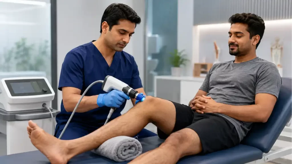
Using pressure waves, this non-invasive procedure stimulates healing in the fascia.
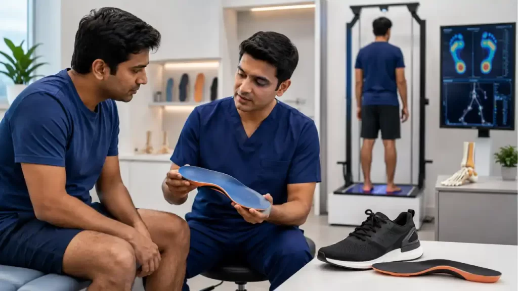
Uses precision-molded inserts that’s designed to rectify biomechanical imbalances and redistribute pressure.
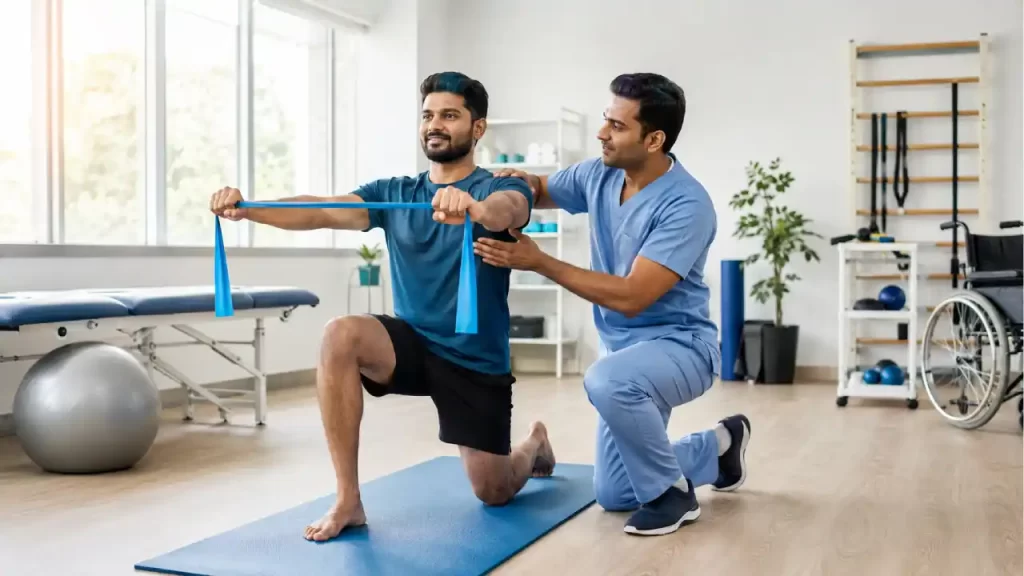
Adopts specific stretching protocols for the Achilles Tendon & Plantar Fascia. They help improve flexibility
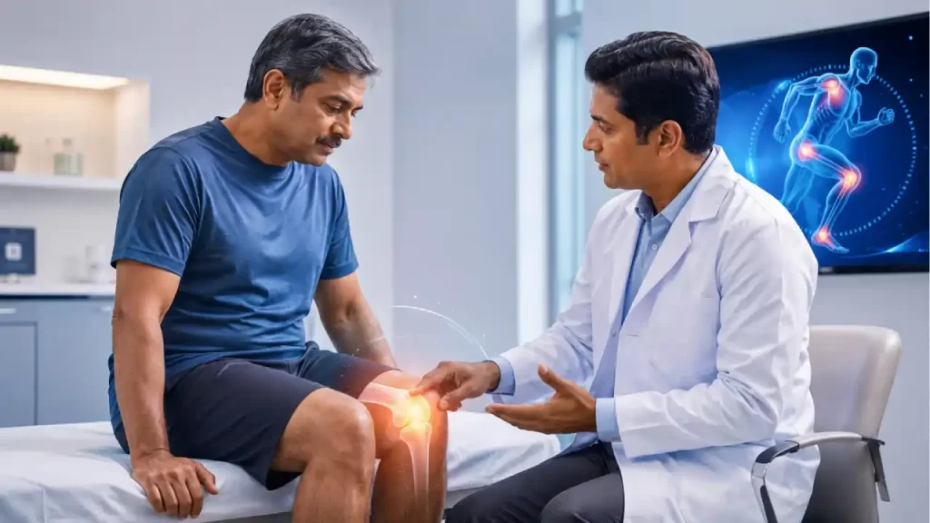
Uses ultrasound-guided injections to drive targeted relief in chronic cases.
The whole process is not easy. A multi-pronged strategy with focus on sustainable recovery and long-term foot health is required to restore your stride.
Start your journey with a virtual consultation to discuss symptoms from home.
We review your medical history and relevant reports for a clear understanding.
Our doctors conduct a thorough assessment through detailed discussions.
We confirm findings with state-of-the-art imaging like X-rays, CT scans, and MRIs.
Our team identifies the root cause and key trigger points for treatment.
We create a customized treatment plan, including necessary medications and procedures.
Our Pain Specialists support a complete recovery focused on total wellness.
We provide ongoing follow-ups tailored to each treatment plan, ensuring continuous care and long-term recovery support.
We relieve your pain, helping you be yourself again!
Spondylosis is an age-related degeneration of the spine’s discs and joints. It happens when the protective cartilage between the vertebrae gradually wears down, leading to stiffness, pain, and reduced spinal flexibility.
Spondylosis pain is mainly caused by wear and tear in the spine. Over time, the discs, joints, and cartilage may degenerate, and bone spurs called osteophytes may develop, causing pain, stiffness, or nerve irritation.
Treatment options may include precision diagnostics, physical rehabilitation, strengthening exercises, spinal alignment therapy, interventional pain management injections, and minimally invasive solutions when required.
Yes. Many cases of spondylosis can be managed with conservative treatment. IBAP Clinics gives priority to non-invasive care such as physical rehabilitation, guided exercises, posture correction, and pain management before considering surgical options.

EXCELLENTTrustindex verifies that the original source of the review is Google. Good pain relief with treatmentPosted on GoogleTrustindex verifies that the original source of the review is Google. Best for pain reliefPosted on GoogleTrustindex verifies that the original source of the review is Google. बहुत अच्छा लगा हमें ये ट्रिटमेंट करा के अच्छे डॉक्टर और अच्छे स्टाफ हैं और काम पैसे में अच्छा अच्छा ट्रिटमेंट होता हैPosted on GoogleTrustindex verifies that the original source of the review is Google. Very nice, good and helpful staf, overall good experiencePosted on GoogleTrustindex verifies that the original source of the review is Google. Very good doctors and good tritment Thank you doctorPosted on GoogleTrustindex verifies that the original source of the review is Google. One of the best clinic for the relief of back painPosted on GoogleTrustindex verifies that the original source of the review is Google. Very good treatment indo british clinic for every pain reliefPosted on GoogleTrustindex verifies that the original source of the review is Google. I have visited indo british pain clinic this is superb clinic for pain relief that's why I have given 5 start rating to this clinic ...Posted on GoogleTrustindex verifies that the original source of the review is Google. My name is Raja Sekhar Reddy Tummuri from kakinada city My father was suffering nervous pain from last 3 years … I have decided to join with this op … Now everything fine after treatment done from DR. vijay sir … Wonderful service received from INDO British advanced pain clinic Hospitals in Hyderabad @ Banjara Hills road no 12.. Each and every staff response , concern , support & everything it’s too good Thank you so much … Special thanks to Mr. DR. Vijay bhaskar Bandi Katla …👏🏻👏🏻👏🏻👏🏻🙏🙏Verified by TrustindexTrustindex verified badge is the Universal Symbol of Trust. Only the greatest companies can get the verified badge who has a review score above 4.5, based on customer reviews over the past 12 months. Read more
Founder & Interventional Pain Specialist — IBAP Clinics, Hyderabad
MBBS · DA · FRCA (London) · FFPMRCA (Pain Medicine, UK) · MBA (Hospital Management)
CCT (Anaesthesia & Pain Medicine, UK) · Advanced Pain Training (Cambridge University Hospitals)
DDSMed Sports Medicine (Chicago) · Fellowship in Neuromodulation & Advanced Pain (London)
Dr Vijay brings over 15 years of postgraduate training across the United Kingdom’s most prestigious institutions — including the Royal College of Anaesthetists, Cambridge University Hospitals, and a dedicated neuromodulation fellowship in London — to his practice in Hyderabad. He is one of very few clinicians in India trained to the level of FFPMRCA — the Faculty of Pain Medicine of the Royal College of Anaesthetists — the highest qualification in pain medicine available in the UK.
His specialist expertise spans the full spectrum of knee pain management: from precision PRP and BMAC injections to cooled radiofrequency genicular nerve ablation, intrathecal drug delivery, and spinal cord stimulation for refractory pain states. He manages cases ranging from the weekend cricketer’s torn meniscus to the elderly cardiac patient with end-stage OA who has been told there are no further options.
Banjara Hills
2nd Floor, 284/A, Road No. 12, above IDFC First Bank, near Omega hospitals, MLA Colony, Banjara Hills, Hyderabad, Telangana 500034.

Indo-British Advanced Pain Clinic © 2026. All Rights Reserved. Designed & Developed By Adroit
WhatsApp us