Helpline :
9807 55 6789
-
Call Us Now: 9807 55 6789Call Us Now: 9807 55 6789
The Condition Salman Khan Called ‘The Disease with the Highest Rates of Suicide’

of TN patients reportsuicidal thoughts(Harvard Medical School, 2025)
pain reliefachieved with Radiofrequency Ablation
long-term cure ratewith MicrovascularDecompression (MVD)
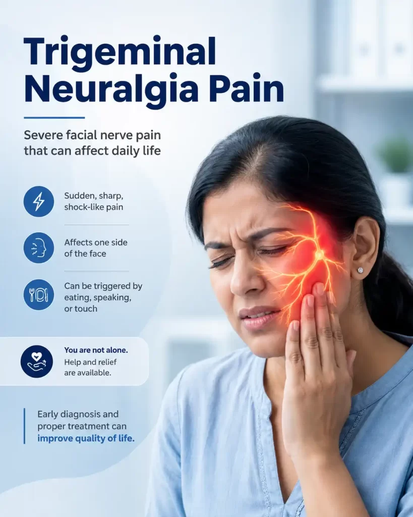
Imagine that the simple act of brushing your teeth sends a lightning bolt of searing electric pain across half your face. Or that a gentle breeze, a sip of cold water, or even the act of speaking triggers an excruciating jolt lasting a few agonising seconds then vanishes as suddenly as it came, leaving you terrified of when the next strike will arrive.
This is the daily reality for people living with Trigeminal Neuralgia (TN) — widely regarded by the medical community as the most severe pain condition known to humanity.
Salman Khan and Trigeminal Neuralgia — Breaking the Silence
Bollywood superstar Salman Khan first experienced TN symptoms whilst filming Partner in 2007. He underwent surgery in the United States in 2011. Speaking whilst promoting Sikandar in Dubai in 2025, he described TN plainly: ‘one disease that basically has the highest rates of suicides.’ He was not exaggerating. A 2025 Swedish study the most comprehensive population study to date confirmed TN as a true suicide disease, associated with increased risk of suicide, depression, and cardiovascular events.
Yet despite this, TN remains widely misdiagnosed often confused with toothache, sinusitis, migraine, or dental problems leaving patients without appropriate treatment for years. Patients are given antidepressants or dental treatments that do not work. Some have had healthy teeth extracted. This article aims to change that.
Why Is It Called the Suicide Disease?
A landmark 2025 study from Harvard Medical School (Fishbein et al., J Pain Research) recruited 229 adults with TN. Results: over 34.6% reported suicidal thoughts in the past two weeks; 57.7% reported thinking about their own death; over 28% had elevated depression. Crucially, this is not psychiatric illness — it is the direct consequence of extreme, relentless, untreatable-feeling pain. The name is not metaphor. It is clinical reality
Think of your face as a large country estate with three postal districts upper, middle, and lower. The trigeminal nerve (the fifth cranial nerve, CN V) is the national postal service responsible for carrying all sensation from the face to the brain.
It has three main branches (divisions), referred to by Roman numerals:
All three branches converge at the Gasserian (trigeminal) ganglion think of it as the main sorting office located deep inside the skull. In TN, this nerve misfires dramatically: instead of calmly relaying sensation, it erupts with extreme, uncontrolled electrical signals — like a faulty live wire sparking without warning.
Neurovascular Conflict — The Most Common Cause (But NOT the Only One)
The most well-understood cause is neurovascular conflict: a blood vessel (most often the Superior Cerebellar Artery) pressing against the trigeminal nerve root where it enters the brainstem.
The Fraying Cable Analogy
Think of the nerve like an electrical cable covered in protective insulation (myelin). Over years, the rhythmic pulsing of an artery against the nerve gradually damages this insulation. Once exposed, the bare wire misfires — creating lightning-bolt pain attacks with no warning. This process is called segmental demyelination.
IMPORTANT: Neurovascular Conflict Is Not Always the Cause — and Surgery Is Not Always the Answer
Not everyone who has a blood vessel touching their trigeminal nerve on MRI will develop TN. Conversely, some patients with classic TN symptoms show no compression at all on imaging. A finding on a scan must always be interpreted in the full clinical context. Surgical decompression is not automatically indicated by an MRI finding alone.
The textbook presentation. Responds best to all treatments.
More treatment-resistant. Constant background pain with or without sharp attacks.
TN is one of the most misdiagnosed conditions in medicine — frequently confused with its closest mimics. The treatment for each condition is entirely different, making accurate diagnosis essential.
The Single Most Important Distinguishing Feature
In TN, patients sit very still during an attack moving the face worsens the pain. In cluster headache (and other TACs), patients cannot sit still they pace, rock, and are visibly agitated. This single observation often clinches the diagnosis at the bedside.
| Feature | TN | Migraine | Dental Pain |
|---|---|---|---|
| Location | Face — jaw, cheek, eye | Hemicranial (one side of head) | Tooth, jaw, localised |
| Duration | Seconds–2 minutes | 4–72 hours | Continuous or aching |
| Light/noise sensitivity | No | Yes — prominent | No |
| Nausea/vomiting | No | Common | Occasional |
| Feather-light skin trigger | Yes — hallmark | No | No |
| Complete pain-free intervals | Yes | Yes | No |
| Response to dental treatment | None | None | Partial |
The Dental Misdiagnosis Trap
TN is one of the most commonly misdiagnosed conditions in dentistry. Patients may undergo unnecessary tooth extractions, root canal treatments, and even sinus operations before TN is recognised. Key distinguishing feature: dental pain does not resolve completely between episodes, and is not triggered by a feather-light touch to the skin whereas TN typically is. If tooth extraction has not relieved the pain, consider a pain specialist referral immediately.
The diagnosis of TN is fundamentally clinical based on a thorough history and examination. There is no blood test for TN. The International Headache Society’s ICHD-3 criteria require:
A skilled pain specialist or neurologist should be able to diagnose classical TN1 from the history alone. Investigations serve to classify the subtype and guide treatment planning.
An ordinary MRI brain scan is insufficient for TN assessment. A dedicated trigeminal MRI protocol is required specifically designed to visualise the fine anatomy of the nerve root entry zone.
| MRI Sequence | Purpose & What It Shows |
|---|---|
| FIESTA / CISS (3D) | Gold standard for TN. Visualises the trigeminal nerve as a bright structure against dark CSF. Identifies vascular loops pressing against the nerve root — neurovascular conflict. |
| 3D TOF MRA | Maps blood vessels near the nerve to identify the specific culprit artery (usually Superior Cerebellar Artery). |
| T1 / T2 / FLAIR (standard) | Excludes secondary causes: tumours, MS plaques, stroke, or other structural lesions. |
| DWI | Excludes acute ischaemic events that may present as acute facial pain. |
Important — A Negative MRI Does NOT Rule Out TN
A negative or inconclusive MRI must never lead a clinician to dismiss a patient’s symptoms. Many patients with classical TN have no vascular contact visible on imaging yet their symptoms, treatment response, and clinical course are entirely consistent with TN. The diagnosis remains clinical. The MRI guides treatment choice, not the diagnosis itself.
The good news: TN is one of the most treatable chronic pain conditions in medicine. The pathway is progressive starting with the simplest interventions and escalating as needed. Surgery is not always required.
Carbamazepine (Tegretol, Mazetol) is an anticonvulsant originally designed to treat epilepsy. In TN, it stabilises sodium channels in nerve membranes, essentially turning down the volume on an overexcited nerve.
The Faulty Car Alarm Analogy
The trigeminal nerve in TN is like a faulty car alarm going off at the slightest disturbance. Carbamazepine is the mechanic who adjusts the sensitivity threshold so only a genuine emergency (actual injury or touch) triggers the alarm, not a gentle breeze or a sip of water.
Efficacy: 70–80% of patients experience significant initial pain relief. This is the only medication with Level A evidence for TN from the European Federation of Neurological Societies (EFNS).
MANDATORY BLOOD TESTS & MONITORING FOR INDIAN PATIENTS Carbamazepine Safety
Carbamazepine is not a drug to take without careful medical supervision. The following monitoring is essential before starting and during treatment:
| Test Required | Why It Matters |
|---|---|
| Full Blood Count (FBC) — before start, then 2-weekly x 3 months, then every 6 months | Carbamazepine can rarely cause agranulocytosis (dangerous drop in white cells) or aplastic anaemia — a potentially fatal condition. |
| Liver Function Tests (LFTs) | Monitors for hepatic effects and drug metabolism. |
| Serum Sodium (Na+) | Carbamazepine commonly causes hyponatraemia (low sodium) — especially dangerous in elderly patients, causing confusion and falls. |
| HLA-B*1502 Genetic Test — CRITICAL IN INDIAN PATIENTS | South and East Asian populations have a higher prevalence of this genetic variant, which dramatically increases risk of Stevens-Johnson Syndrome (SJS) / Toxic Epidermal Necrolysis — potentially fatal skin reactions. At IBAP Clinics, we routinely perform this test before initiation. |
| Drug | Mechanism | Role in TN |
|---|---|---|
| Oxcarbazepine (Trileptal) | Sodium channel stabiliser (similar to carbamazepine, cleaner profile) | Often preferred first-line in India — less blood monitoring required, better tolerated. Check sodium levels. |
| Gabapentin | Calcium channel modulator | Adjunct therapy or monotherapy when carbamazepine intolerable. Better for TN2 (constant burning component). |
| Pregabalin | Calcium channel modulator | Similar to gabapentin; TN2, MS-related TN, atypical facial pain. |
| Baclofen | GABA-B receptor agonist | Useful as add-on therapy. Evidence for TN1 as adjunct. |
| Lamotrigine | Sodium/calcium channel modulator | Adjunct to carbamazepine. Very slow titration required to avoid rash. |
| Amitriptyline | Tricyclic antidepressant (TCA) | More useful in TN2 with constant background aching — NOT effective for classical TN1 attacks. |
| IV Lidocaine infusions | Sodium channel blockade | Bridging therapy during acute severe episodes or medication transitions — hospital-based. |
An Important Clinical Reality — Medication Efficacy Fades Over Time
Many patients initially respond well to medication, but drug efficacy tends to diminish as the disease progresses. Side effects also accumulate with higher doses. This is the typical juncture at which interventional options become appropriate — and should not be delayed unnecessarily.
All percutaneous (needle-based) procedures target the Gasserian ganglion the central sorting office of the trigeminal nerve via a needle passed through a small natural opening in the skull called the foramen ovale, under X-ray fluoroscopy guidance.
A fine, insulated needle is advanced through the foramen ovale to the Gasserian ganglion. Electrical stimulation confirms correct position the patient feels tingling in exactly the distribution of their pain. Heat is then applied (60–80°C for 60–90 seconds), selectively damaging the pain fibres whilst attempting to spare the touch fibres.
The Selective Wire-Burning Analogy
Imagine a bundle of wires causing a short circuit. RFA is the electrician who selectively burns only the faulty wires, leaving the rest of the cable functional. The pain fibres are damaged, whilst larger touch fibres are largely preserved.
A catheter with a small balloon at its tip is advanced through the foramen ovale. The balloon is inflated inside Meckel’s cave (a small pocket around the Gasserian ganglion), applying mechanical pressure to the nerve. This damages pain fibres whilst tending to preserve corneal touch fibres better than RFA.
The Pause Button Analogy
If RFA is the electrician burning wires selectively, balloon compression is a gentle squeeze that disrupts the signal pathway physically like pressing the pause button rather than cutting wires. Because it spares corneal fibres, it is particularly preferred for V1 (eye/forehead) TN.
Procedure: Performed under brief general anaesthesia (typically under 30 minutes). Excellent for V1 TN and multi-branch involvement. Key side effect: temporary facial numbness and chewing difficulty. Bradycardia during balloon inflation is monitored by the anaesthetic team.
A neurosurgical procedure under general anaesthesia. A small opening (craniotomy) is made behind the ear. The trigeminal nerve is visualised under a microscope, and the offending blood vessel is carefully separated from the nerve using a small Teflon sponge or padding.
The Maintenance Engineer Analogy
MVD is like the maintenance engineer who discovers an electrical cable has been chafed by a pipe for years. Rather than cutting the cable (ablation), the engineer inserts padding between the pipe and cable removing the source of irritation and allowing the cable to heal naturally. MVD is the only procedure that corrects the underlying cause rather than disrupting the pain pathway.
Prerequisite: Confirmed vascular contact on MRI trigeminal protocol is strongly recommended before MVD is offered. MVD where no vascular conflict is found intraoperatively has a lower success rate. Long-term pain-free rates of 70–80% at 5 years are the best of any procedure for classical TN1 with confirmed neurovascular conflict.
A completely non-invasive option for patients unfit for any needle or open procedure. Over 200 highly focused beams of radiation are precisely directed at the trigeminal nerve root entry zone. No incision, no needle, no general anaesthesia.
The 200 Torchbeams Analogy
Imagine 200 weak torch beams, each harmless on its own, all focused to converge at a single point. At that convergence, the energy is sufficient to damage the nerve selectively without touching anything else in the path. Pain relief typically takes 4–8 weeks to develop, unlike the immediate relief from RFA or MVD.
Ideal for: Elderly, very frail patients; those on anticoagulants (blood thinners); those who have failed other interventions; patients who refuse any invasive procedure. Available at select major centres in Hyderabad including Apollo Hospitals.
Outcomes: Approximately 60–70% pain-free at 2 years. Slightly inferior long-term outcomes compared to MVD, but the lowest procedural risk of any invasive option.
| Parameter | Radiofrequency Ablation (RFA) | Microvascular Decompression (MVD) |
|---|---|---|
| Mechanism | Nerve ablation — destroys pain fibres (destructive) | Nerve-preserving — removes vascular pressure (non-destructive) |
| Anaesthesia | Local + light IV sedation | General anaesthesia + craniotomy |
| Hospital stay | Day procedure / 1 night maximum | 3–5 days inpatient |
| Immediate pain relief | ~85–90% | ~90–95% |
| Pain-free at 5 years | 50–60% | 70–80% |
| Recurrence risk | 20–30% at 5 years | 10–20% at 5 years |
| Facial numbness | Expected — common side effect | Minimal to none |
| Serious complication risk | Low — anaesthesia dolorosa ~2–5% | Higher — stroke, hearing loss, CSF leak (rare but real) |
| MRI NVC required | No — effective even without NVC | Yes — strongly preferred prerequisite |
| Nerve preserved? | No | Yes |
| Repeatability | Yes — can be repeated | Generally a one-time procedure |
| Ideal patient profile | Elderly, frail, medically unfit, MS-related TN, no NVC | Young, fit, confirmed NVC on MRI, willing to accept surgical risk |
| Availability in India | Widely available at specialist pain centres | Major neurosurgical centres only |
The Clinical Bottom Line
For a fit 45-year-old with MRI-confirmed vascular compression, MVD is the gold standard best long-term cure rates and nerve preservation. For an 80-year-old with significant medical comorbidities and the same diagnosis, RFA is the wiser, safer choice. Neither is universally superior. The right answer is always patient-specific determined by age, fitness, MRI findings, patient preference, and the surgeon’s or interventionalist’s experience.
TN is not a psychiatric condition but it profoundly affects mental health. Research shows a significantly higher risk of newly diagnosed depression, anxiety disorders, and sleep disturbances following TN diagnosis largely due to its devastating impact on psychological, behavioural, and social functioning.
Suicidality is an urgent yet under-addressed concern. It is associated not with psychiatric illness per se, but with high levels of anxiety, depression, and pain intensity. Patients are often reluctant to discuss the psychological impact of their pain particularly in India, where mental health discussions carry stigma.
At IBAP Clinics, our pain management approach is holistic: we treat the pain and address the psychological burden, working collaboratively with psychiatry and psychology as needed.
Crisis Support — If You Are Struggling
If you or a loved one are experiencing suicidal thoughts due to chronic pain, please reach out. iCall (India): 9152987821. Vandrevala Foundation: 1860-2662-345. You are not alone, and effective treatment exists.
Start your journey with a virtual consultation to discuss symptoms from home.
We review your medical history and relevant reports for a clear understanding.
Our doctors conduct a thorough assessment through detailed discussions.
We confirm findings with state-of-the-art imaging like X-rays, CT scans, and MRIs.
Our team identifies the root cause and key trigger points for treatment.
We create a customized treatment plan, including necessary medications and procedures.
Our Pain Specialists support a complete recovery focused on total wellness.
We provide ongoing follow-ups tailored to each treatment plan, ensuring continuous care and long-term recovery support.
We relieve your pain, helping you be yourself again!
Trigeminal neuralgia (TN) is a chronic pain condition of the fifth cranial nerve causing sudden, intense electric-shock pain in the face, jaw, or eye. It is called the suicide disease because studies show over one-third of TN patients report suicidal thoughts not from psychiatric illness, but because the pain is so extreme and often unrecognised. Bollywood star Salman Khan has publicly described his battle with TN.
TN causes brief (seconds), electric-shock facial pain triggered by touch or eating, with no autonomic symptoms. Cluster headaches cause longer (15 min–3 hrs) boring orbital pain with tearing, red eye, and nasal congestion and patients cannot sit still. Migraines cause throbbing head pain lasting hours, often with nausea and light sensitivity. Treatment for all three is completely different.
No. Most patients are initially managed successfully with medications (carbamazepine or oxcarbazepine). When drugs fail, minimally invasive day procedures such as radiofrequency ablation (RFA) achieve excellent relief without open surgery. Microvascular decompression (MVD) surgery is reserved for suitable younger, fitter patients with confirmed vascular compression on MRI.
A standard MRI brain scan cannot resolve the fine anatomy of the trigeminal nerve root. A dedicated trigeminal protocol uses FIESTA or CISS sequences — showing the nerve and adjacent blood vessels in high definition to identify neurovascular conflict. This finding directly guides treatment decisions, particularly regarding microvascular decompression surgery.
Carbamazepine requires careful monitoring. Key concerns in Indian patients: (1) regular blood counts to detect rare blood cell reduction; (2) liver function and sodium checks; and (3) HLA-B*1502 genetic testing — this variant is more prevalent in South and East Asian populations and significantly increases risk of Stevens–Johnson Syndrome, a potentially fatal skin reaction. At IBAP Clinics, we screen for this routinely before starting treatment
RFA involves passing a fine needle to the trigeminal ganglion and applying controlled heat to selectively damage pain-carrying nerve fibres. It is performed as a day procedure under local anaesthesia with light sedation no general anaesthesia required. This makes it ideal for elderly or medically unfit patients. Success rates are approximately 85–90%, with some expected facial numbness.
At IBAP Clinics (Banjara Hills and Hafeezpet, Hyderabad), Dr Vijay Bhaskar Bandikatla offers a complete, individualised TN pathway including specialist MRI review, evidence-based medication with appropriate safety monitoring including HLA-B*1502 screening, and the full range of interventional procedures. Appropriate neurosurgical referral pathways are in place for MVD and Gamma Knife when indicated.

EXCELLENTTrustindex verifies that the original source of the review is Google. Good pain relief with treatmentPosted on GoogleTrustindex verifies that the original source of the review is Google. Best for pain reliefPosted on GoogleTrustindex verifies that the original source of the review is Google. बहुत अच्छा लगा हमें ये ट्रिटमेंट करा के अच्छे डॉक्टर और अच्छे स्टाफ हैं और काम पैसे में अच्छा अच्छा ट्रिटमेंट होता हैPosted on GoogleTrustindex verifies that the original source of the review is Google. Very nice, good and helpful staf, overall good experiencePosted on GoogleTrustindex verifies that the original source of the review is Google. Very good doctors and good tritment Thank you doctorPosted on GoogleTrustindex verifies that the original source of the review is Google. One of the best clinic for the relief of back painPosted on GoogleTrustindex verifies that the original source of the review is Google. Very good treatment indo british clinic for every pain reliefPosted on GoogleTrustindex verifies that the original source of the review is Google. I have visited indo british pain clinic this is superb clinic for pain relief that's why I have given 5 start rating to this clinic ...Posted on GoogleTrustindex verifies that the original source of the review is Google. My name is Raja Sekhar Reddy Tummuri from kakinada city My father was suffering nervous pain from last 3 years … I have decided to join with this op … Now everything fine after treatment done from DR. vijay sir … Wonderful service received from INDO British advanced pain clinic Hospitals in Hyderabad @ Banjara Hills road no 12.. Each and every staff response , concern , support & everything it’s too good Thank you so much … Special thanks to Mr. DR. Vijay bhaskar Bandi Katla …👏🏻👏🏻👏🏻👏🏻🙏🙏Verified by TrustindexTrustindex verified badge is the Universal Symbol of Trust. Only the greatest companies can get the verified badge who has a review score above 4.5, based on customer reviews over the past 12 months. Read more

MBBS, DA, FRCA (UK), FFPMRCA (Pain Medicine, RCOA, UK)
CCT (Anesthesiology And Pain Management)
Neuromodulation & Advanced Pain Research Fellowship (London), MBA (HM)
Banjara Hills
2nd Floor, 284/A, Road No. 12, above IDFC First Bank, near Omega hospitals, MLA Colony, Banjara Hills, Hyderabad, Telangana 500034.

Indo-British Advanced Pain Clinic © 2026. All Rights Reserved. Designed & Developed By Adroit
WhatsApp us