Helpline :
9807 55 6789
- Call Us Now: 9807 55 6789Call Us Now: 9807 55 6789
Indians estimated to live with osteoporosis
Women over 50 will suffer an osteoporotic fracture
T-score threshold for osteoporosis on DEXA scan
Of vertebral fractures are initially misdiagnosed or missed
Of osteoporosis risk is modifiable — through diet, sun, and exercise
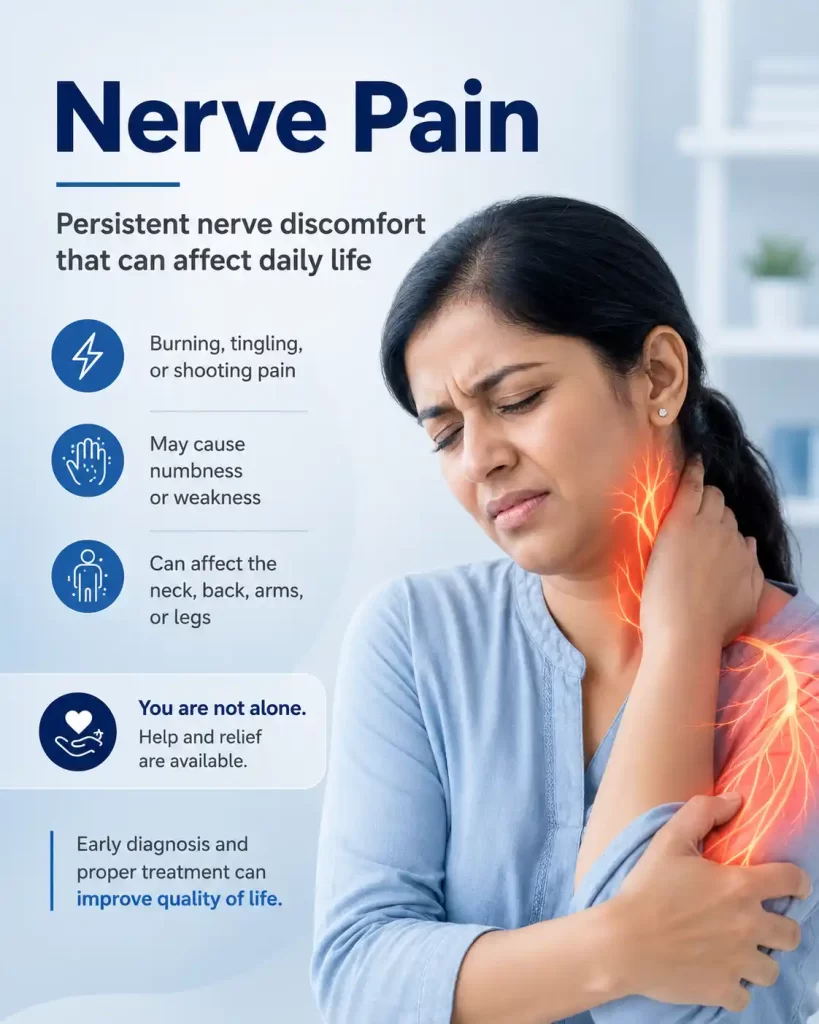
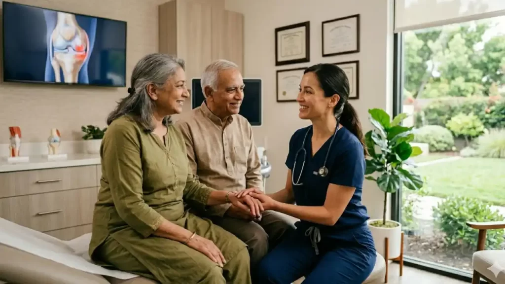
There is a particular kind of pain I see again and again in my clinic. A 65-year-old lady — let us call her Usharani — bends forward to pick up her grandchild and feels a sudden, searing crack in her middle back. She spends three days telling herself it is just a muscle pull. By the time she comes to see me, she cannot sit upright for more than a few minutes. Her X-ray reveals a collapsed vertebra. Her spine — the very scaffolding that has held her upright through decades of cooking, carrying, and caring — has silently crumbled from within.
This is osteoporosis. Not just thinning bones on a scan. Real pain. Real disability. Real fear. And — and this is what I want every patient who reads this to understand — real, treatable suffering.
As a pain physician, I see osteoporosis from a different vantage point than an orthopaedic surgeon or a general physician. My interest is not just in what has broken, but in why it hurts, how severely it is threatening the nervous system, and what we can do — right now — to restore dignity and function to patients whose pain has been dismissed, underestimated, or simply untreated for far too long.
Think of healthy bone like reinforced concrete — steel rods (the protein matrix, mostly collagen) embedded in a hard mineral matrix (calcium hydroxyapatite). The two components work together. The protein gives flexibility and tensile strength; the calcium gives hardness and compressive resistance. Remove either one, and the structure weakens catastrophically.
In osteoporosis, both are depleted. The bone becomes less dense, less strong, less resilient. Microscopically, the spongy inner bone — the trabecular bone — loses its honeycomb lattice and becomes more like a sparse web. The hard outer shell — the cortical bone — thins. What was once a robust girder becomes something closer to hollow chalk.
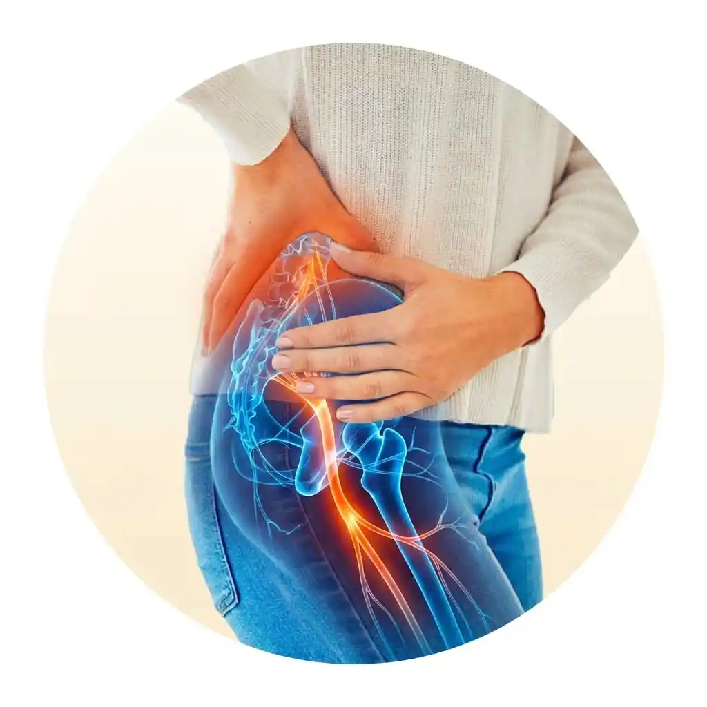
And here is the cruel part: it happens silently. No warning. No pain from the bone loss itself. Until the day a vertebra collapses, a hip fractures, or a wrist snaps — often from forces as modest as a cough, a sneeze, or bending over. We call these fragility fractures, and they are among the most underrecognised sources of chronic pain in India today.
Osteoporosis is a "silent disease" until a fracture occurs — but once fractures begin, the risk of subsequent fractures rises dramatically. One vertebral fracture increases the risk of a second fracture by 5-fold within the next 12 months. Early diagnosis and treatment are not optional. They are urgent.
Bone is not static. It is living tissue, constantly being broken down by cells called osteoclasts and rebuilt by cells called osteoblasts. In youth, building outpaces breakdown. In osteoporosis, this balance tips — sometimes dramatically — towards destruction.
Every one of these causes is either preventable, treatable, or both. The tragedy is that most patients do not know their bones are at risk until something breaks.
There is something I say to my patients that surprises them every time — and it needs to be said openly. India has a deep-rooted cultural obsession with lighter skin. It runs through matrimonial ads, film posters, and cosmetics counters alike. Millions of Indian women — and increasingly men — actively avoid sun exposure to prevent tanning, and apply fairness creams, skin-lightening products, and high-SPF sunscreens as part of their daily routine, often from teenage years onwards.
The painful irony is this: the very habits that protect skin colour are quietly stripping calcium from bones. This is not a minor effect. The consequences are measurable, irreversible, and arriving decades later in the form of fractures.
Many women deliberately stay indoors, use umbrellas, and wear full-sleeve clothing — not for health, but to stay fair. Vitamin D synthesis requires direct UV-B contact with unprotected skin. There is no workaround.
SPF 30+ sunscreen can reduce vitamin D skin synthesis by up to 95%. Applied every morning before stepping out, it effectively eliminates the body's primary vitamin D production mechanism.
Skin-lightening products containing titanium dioxide, zinc oxide, or kojic acid create a physical UV barrier on the skin. Used daily, they have a similar sun-blocking effect — blocking the UV-B that the skin needs to produce vitamin D.
Fair-complexioned, health-conscious women in their 50s — many of whom have eaten well, exercised, and taken care of themselves — arrive with T-scores of −3.0 or worse. They are stunned. When I ask about their sun habits and skincare routine, the picture becomes clear immediately. They have diligently avoided the sun their entire adult lives. The fairness came at a price nobody warned them about.
Think of your bones like a savings account. From birth to around age 30, you are depositing — building bone mass. From your 30s onwards, small withdrawals begin. At menopause or with illness, the withdrawals accelerate. Osteoporosis is when the account goes deeply into the red — and the bank (your spine) starts bouncing cheques (fractures).
— Dr Vijay Bandikatla, IBAP ClinicsOsteoporosis: Bone Loss, Fracture Cascade & Intervention Points

When a patient walks into my pain clinic with sudden-onset back pain — particularly an elderly woman, or anyone on long-term steroids — my first thought is a vertebral compression fracture. The workup is systematic. Every investigation answers one specific clinical question. Here is exactly what I order, and why.
📊 What Does My T-Score Mean?
DEXA does not diagnose a current fracture — X-ray and MRI do that. DEXA predicts your future fracture risk.
A T-score alone does not tell the full story. Two people can have the same T-score but very different fracture risks. That is why I use the FRAX tool — developed by the World Health Organisation — alongside DEXA. It calculates your personal 10-year probability of a major fracture by factoring in:
A T-score without FRAX is like a blood pressure reading without knowing the patient's age, diabetes status, and family history. The number alone is not enough. A 75-year-old woman with a T-score of −2.0, a prior wrist fracture, and three years of steroid use is in a completely different risk category than a 52-year-old with the same T-score and no other risk factors. Same number. Very different decisions. That is what FRAX does — it gives context to the number.
— Dr Vijay Bandikatla, IBAP ClinicsHere is a test that I think is genuinely underutilised in India, and one that can make a real difference to early detection. The heel quantitative ultrasound scan (QUS) measures how sound waves travel through the heel bone (calcaneus). It produces a stiffness index and a T-score equivalent — without any radiation.
If you are in any of these groups — postmenopausal woman · long-term steroid user · chronic vitamin D deficiency · rheumatoid arthritis · prior fragility fracture — a normal QUS result is not a reason to relax. These patients must proceed to a formal DEXA scan regardless. The QUS opens the door. DEXA tells you what is truly behind it.
I routinely request: serum calcium and phosphate; 25-hydroxyvitamin D; parathyroid hormone (PTH); thyroid function; full blood count; renal and liver function; bone turnover markers such as serum CTX (C-terminal telopeptide) and P1NP (procollagen type I N-propeptide). In any patient with atypical features, I add serum protein electrophoresis to exclude myeloma. These tests serve two purposes: they identify treatable secondary causes, and they help me monitor the response to treatment over time.
Evidence Table| Investigation | What It Shows | Clinical Question Answered | Limitation |
|---|---|---|---|
| Plain X-Ray | Vertebral height loss, wedge/biconcave deformity | Is there a fracture? Which level? | Misses soft tissue, cord, acute vs chronic |
| CT Scan | Cortical integrity, retropulsed fragments, canal narrowing | Is the posterior wall breached? Safe for cement? | Radiation; poor soft tissue contrast |
| MRI Spine | Cord compression, oedema, malignant vs benign, ligaments | Is the nervous system at risk? Acute or chronic? | Cost; availability; contraindicated with some implants |
| Calcaneal QUS Screening | Heel bone stiffness index; T-score equivalent; fracture risk stratification | Is this patient at elevated risk? Should they proceed to DEXA? | Not a DEXA substitute; heel only; affected by soft tissue; not for monitoring treatment |
| DEXA Scan Gold Standard | BMD at lumbar spine, hip, forearm; T-score and Z-score; FRAX 10-year fracture risk | Confirm osteoporosis diagnosis; quantify fracture risk; monitor treatment? | Requires dedicated machine; radiation (low); not available universally; cost |
| Blood Tests | Calcium, VitD, PTH, bone turnover markers, myeloma screen | Secondary cause? Severity? Response to Rx? | Multiple tests needed; interpretation requires expertise |
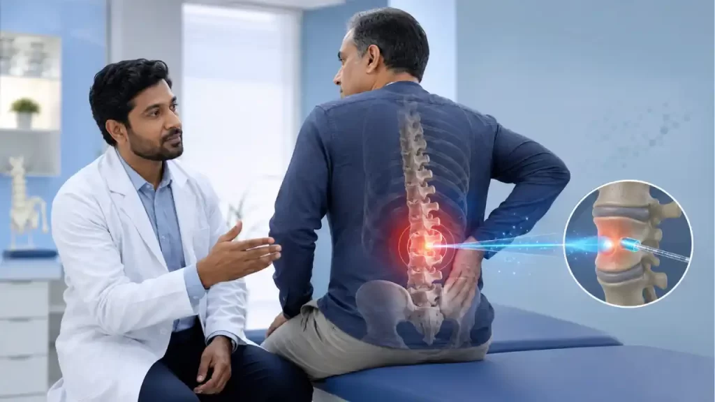
I want to spend a moment on this, because the pain of a vertebral compression fracture is still frequently dismissed. Patients are told, "At your age, back pain is normal." It is not. Acute severe back pain in an elderly person — especially a postmenopausal woman, or someone on steroids — is an osteoporotic fracture until proven otherwise.
The pain is typically acute onset, localised to the mid or lower back, worsened dramatically by standing and loading, and partially relieved by lying flat. Breathing deeply can be exquisite agony. Rolling over in bed. Standing from a chair. The patient cannot find a comfortable position.
When a fracture is at the thoracolumbar junction (T12-L2, the most common site) and a fragment or collapsed body encroaches on the spinal canal, radicular pain appears — shooting down the buttocks, thighs, or even the feet. In severe cases, cauda equina syndrome with bladder and bowel dysfunction represents a surgical emergency. These patients do not belong at home resting; they need urgent MRI and neurosurgical assessment.
If a patient with a known or suspected vertebral fracture develops: new leg weakness or numbness; difficulty passing urine or opening bowels; saddle anaesthesia (numbness in the inner thighs and perineum); or rapidly progressive pain — these are cord compression red flags. Do NOT wait. An urgent MRI and neurosurgical consult are needed within hours, not days.
Here is where my perspective diverges from what many patients expect. Most people assume osteoporosis means calcium tablets and maybe a drug from the GP. When a fracture has occurred, the management must be active, multimodal, and sequenced. Let me walk you through what we actually do.
A small amount of rest — one to two weeks maximum — is reasonable in the acute phase. Semi-recumbent, firm mattress, supported posture. After that, gradual mobilisation with a well-fitted thoracolumbar brace begins. What is never acceptable — and what I see causing serious harm — is insisting that an elderly woman with a vertebral fracture remain completely bed-bound for weeks. The family means well. But prolonged immobility is a second injury.
Bed Rest Cascade Visual🚨 What Prolonged Bed Rest Does to an Elderly Osteoporotic Patient
Each complication feeds the next. This is not a recovery — it is a cascade of decline.
These procedures stabilise the fracture, eliminate the mechanical pain, and allow the patient to sit up, stand, and walk — often within 24 to 48 hours. They break the bed-rest cascade before it starts. In elderly women with osteoporotic vertebral fractures, vertebroplasty and kyphoplasty are not elective comfort measures. They are urgent interventions that prevent DVT, PE, pressure sores, muscle loss, and the spiral into dependency.
Pain management in the acute phase requires a WHO-ladder approach. Regular paracetamol (with adequate dosing — not the subtherapeutic "one tablet if needed" that many patients have been told). NSAIDs with gastroprotection in those without contraindications. Short-term weak opioids — tramadol or codeine — for breakthrough pain. Neuropathic agents (pregabalin, duloxetine) when there is a radicular component. I am cautious with opioids in the elderly — constipation, confusion, and falls from sedation can create more fractures than they prevent. The goal is functional analgesia: pain controlled enough to mobilise and participate in physiotherapy.
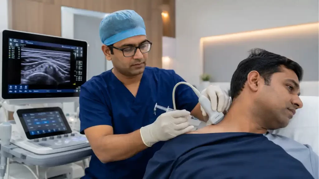
When a collapsed vertebra or retropulsed fragment is irritating or compressing a nerve root, the resulting radicular pain — sharp, electric, shooting — can be truly disabling. An epidural steroid injection (ESI), performed under fluoroscopic guidance, delivers corticosteroid (typically triamcinolone or methylprednisolone) into the epidural space adjacent to the compressed root. This reduces neurogenic inflammation, interrupts the pain signal, and allows meaningful rehabilitation. I have seen patients who were bed-bound and tearful from radicular pain walk out of the procedure room with dramatically reduced symptoms. The ESI is not curative — the mechanical problem persists — but it buys time, improves function, and can be the difference between a patient engaging with rehabilitation versus spiralling into chronic disability.
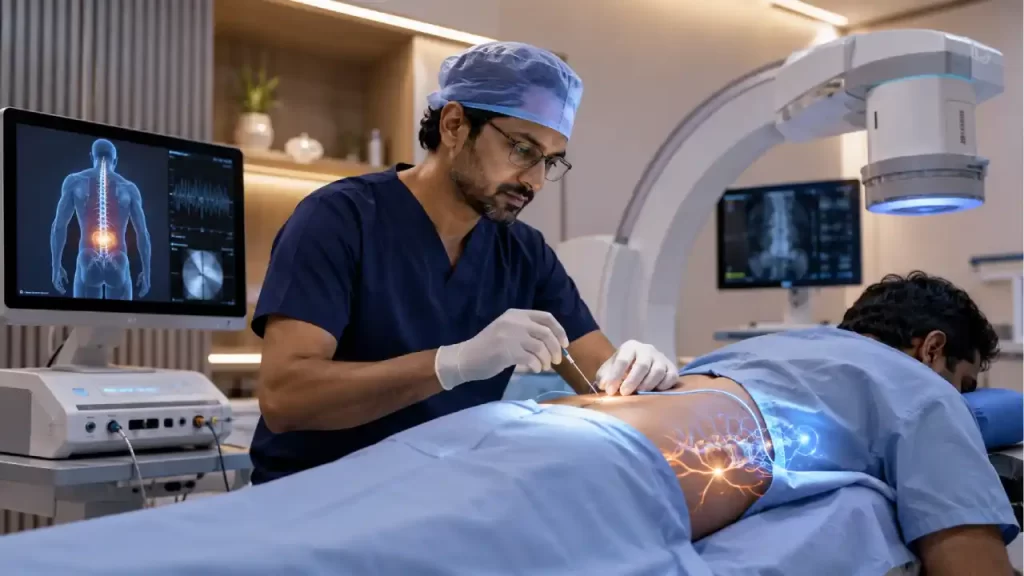
Vertebroplasty is the simpler of the two cement augmentation procedures. Under fluoroscopic (X-ray) and sometimes CT guidance, I pass a trocar — a thick, hollow needle — through the pedicle of the fractured vertebra. Bone cement (polymethylmethacrylate, PMMA) is then injected under pressure directly into the collapsed body, filling the fracture clefts and trabecular spaces. As the cement cures (polymerises), it generates heat and stabilises the fracture. The pain relief, in carefully selected patients, can be dramatic — often within 24 to 48 hours. The procedure takes 45–60 minutes, is performed under sedation or light general anaesthesia, and patients typically go home the next day.
Kyphoplasty adds an important step before cement injection. After positioning the trocar, a deflated balloon (balloon tamp) is inserted into the vertebral body and inflated under controlled pressure. This balloon compacts the surrounding bone, creates a cavity, and — crucially — can partially restore the lost vertebral height and reduce the kyphotic (forward-bending) deformity. The balloon is then deflated and removed, and cement is injected into the cavity at lower pressure, reducing the risk of cement leakage compared to vertebroplasty. Kyphoplasty is preferable in more acute fractures (where height restoration is possible), in patients with significant kyphotic deformity, and when the posterior cortex is partially compromised (the lower injection pressure is safer).
Comparison Table| Feature | Vertebroplasty | Kyphoplasty |
|---|---|---|
| Balloon Used | No | Yes |
| Height Restoration | Minimal | Partial — often significant |
| Cement Injection Pressure | Higher | Lower (into pre-formed cavity) |
| Cement Leakage Risk | Higher (~10–15%) | Lower (~5–9%) |
| Kyphosis Correction | Minimal | Better |
| Procedure Duration | ~45 minutes | ~60–75 minutes |
| Best Indication | Acute/subacute fracture, stable walls | Significant collapse, kyphosis, or compromised posterior wall |
| Pain Relief Efficacy | Excellent | Excellent |
When vertebroplasty or kyphoplasty cannot do enough — because of significant spinal instability, progressive neurological deficit, or multilevel disease — spine surgery with pedicle screw fixation becomes necessary. But I want families to understand two important realities that do not always get explained clearly.
Surgery Risk Box🏥 Why Surgery Is High-Risk in the Elderly
🔩 Why Screws Fail in Osteoporotic Bone
🌍 The Access Gap in India — A Harder Truth
For many years — and still today outside major cities — vertebroplasty and kyphoplasty were simply not available to most patients who needed them. Not because they did not qualify. Because the trained operators, fluoroscopy suites, and cement systems were concentrated in a handful of tertiary centres. Women in tier-2 cities were prescribed bed rest, analgesics, and calcium tablets — not because that was the best treatment, but because nothing better was on offer. This is changing. Slowly. Which is why I feel strongly about making these procedures accessible and ensuring patients and families know to ask for them.
Trying to fix a screw into osteoporotic bone without cement augmentation is like trying to hang a heavy curtain rail with rawlplugs in crumbling plaster. The wall looks solid from outside. But the anchors have nothing to grip. The rail holds for a week, then falls at 3 am. Cement augmentation is the upgrade to solid masonry fixings — and in osteoporotic spine surgery, it is not optional.
— Dr Vijay Bandikatla, IBAP ClinicsHere is the truth that most patients do not hear until it is too late: osteoporosis is largely preventable. And even after a fracture, the trajectory can be slowed, halted, and sometimes partially reversed. Prevention is not a single act — it is a set of daily habits that accumulate over decades. Think of it as a savings account you start contributing to from your 20s, knowing you will need those reserves in your 60s and 70s.
Prevention Pillars GridYou do not need expensive supplements or imported foods to protect your bones. The Indian kitchen — when used well — is one of the richest sources of bone-building nutrition in the world. The problem is that most people are not eating enough of these foods, or are preparing them in ways that reduce their nutritional value. Here is what I recommend to my patients every day.
Indian Foods SVG InfographicBone-Building Foods from the Indian Kitchen — Calcium & Key Nutrients

🌅 Morning Habits
🍽️ Meal Additions
🚶 Movement Every Day
Building bone health is like filling a water tank. You spend your 20s and 30s filling it to maximum. From your 40s, small leaks begin. Menopause, steroid use, and poor diet tear large holes in the tank. By the time it empties — that is the fracture. The best time to start filling it was 30 years ago. The second best time is today.
— Dr Vijay Bandikatla, IBAP ClinicsTreating the fracture and its pain without simultaneously treating the osteoporosis is like patching a leak without turning off the tap. Every patient who leaves my clinic with a vertebral fracture diagnosis gets a concurrent conversation about bone health optimisation.
Calcium supplementation (1,000–1,200 mg daily in divided doses, with meals); vitamin D (cholecalciferol, typically 2,000–4,000 IU daily in deficient Indian patients); protein optimisation through diet counselling; and where appropriate, referral to an endocrinologist or rheumatologist for anti-resorptive or anabolic therapy. Bisphosphonates (alendronate, zoledronic acid) remain the cornerstone of pharmacological treatment, with denosumab, teriparatide, and romosozumab for high-risk or refractory cases. These decisions require specialist collaboration and cannot be made in isolation.
I want to close the clinical section with something that is not in any guideline but matters enormously: the way we listen to these patients.
Most of them are elderly women. Many come with family members who speak over them, summarise their symptoms for them, or minimise what they are experiencing. "She is just dramatic," or "At her age, some pain is expected." I have heard versions of this in my consultation room more times than I can count.
Here is what I know from 15 years of pain medicine: undertreated pain is not a minor inconvenience. It triggers the hypothalamic-pituitary-adrenal axis. It elevates cortisol chronically, which — circularly — accelerates bone loss. It disrupts sleep, which impairs healing. It causes depression and social withdrawal. It leads to immobility, which causes more bone loss. Untreated pain from an osteoporotic fracture is not just unkind. It is clinically counterproductive.
Understanding and empathy are not soft extras in medical care. They are active ingredients in recovery. Patients who feel heard comply better with treatment, mobilise earlier, and report less chronic pain. If there is one thing I want every family member reading this to take home, it is this: the pain your mother or grandmother is describing is real, it is measurable, and it deserves expert attention — not dismissal.
FAQ AccordionDo not wait. An urgent consultation can determine whether you have a vertebral fracture — and what can be done, right now, to relieve your pain safely and effectively.
Indo British Advanced Pain Clinic
2nd Floor, 284/A, Road No. 12
Above IDFC First Bank, near Omega Hospitals
MLA Colony, Banjara Hills
Hyderabad, Telangana 500034
Indo British Advanced Pain Clinic
Sy No. 2, 4th Floor, Plot No. 200
Beside South India Shopping Mall
Opp. Fortune Heights, Mythri Nagar
Madeenaguda, Hyderabad, Telangana 500049
This article is intended for general educational and informational purposes only. It does not constitute medical advice, diagnosis, or treatment. The content reflects the professional opinion and clinical experience of Dr Vijay Bhaskar Bandikatla and is not a substitute for individualised consultation with a qualified medical professional. Treatments described (including vertebroplasty, kyphoplasty, and epidural steroid injections) carry risks and benefits that must be assessed on a case-by-case basis. Always consult a specialist before making any medical decisions. IBAP Clinics, Hyderabad.
Indians estimated to live with osteoporosis
Women over 50 will suffer an osteoporotic fracture
T-score threshold for osteoporosis on DEXA scan
Of vertebral fractures are initially misdiagnosed or missed
Of osteoporosis risk is modifiable — through diet, sun, and exercise
There is a particular kind of pain I see again and again in my clinic. A 65-year-old lady — let us call her Usharani — bends forward to pick up her grandchild and feels a sudden, searing crack in her middle back. She spends three days telling herself it is just a muscle pull. By the time she comes to see me, she cannot sit upright for more than a few minutes. Her X-ray reveals a collapsed vertebra. Her spine — the very scaffolding that has held her upright through decades of cooking, carrying, and caring — has silently crumbled from within.
This is osteoporosis. Not just thinning bones on a scan. Real pain. Real disability. Real fear. And — and this is what I want every patient who reads this to understand — real, treatable suffering.
As a pain physician, I see osteoporosis from a different vantage point than an orthopaedic surgeon or a general physician. My interest is not just in what has broken, but in why it hurts, how severely it is threatening the nervous system, and what we can do — right now — to restore dignity and function to patients whose pain has been dismissed, underestimated, or simply untreated for far too long.
Think of healthy bone like reinforced concrete — steel rods (the protein matrix, mostly collagen) embedded in a hard mineral matrix (calcium hydroxyapatite). The two components work together. The protein gives flexibility and tensile strength; the calcium gives hardness and compressive resistance. Remove either one, and the structure weakens catastrophically.
In osteoporosis, both are depleted. The bone becomes less dense, less strong, less resilient. Microscopically, the spongy inner bone — the trabecular bone — loses its honeycomb lattice and becomes more like a sparse web. The hard outer shell — the cortical bone — thins. What was once a robust girder becomes something closer to hollow chalk.
And here is the cruel part: it happens silently. No warning. No pain from the bone loss itself. Until the day a vertebra collapses, a hip fractures, or a wrist snaps — often from forces as modest as a cough, a sneeze, or bending over. We call these fragility fractures, and they are among the most underrecognised sources of chronic pain in India today.
Osteoporosis is a "silent disease" until a fracture occurs — but once fractures begin, the risk of subsequent fractures rises dramatically. One vertebral fracture increases the risk of a second fracture by 5-fold within the next 12 months. Early diagnosis and treatment are not optional. They are urgent.
Bone is not static. It is living tissue, constantly being broken down by cells called osteoclasts and rebuilt by cells called osteoblasts. In youth, building outpaces breakdown. In osteoporosis, this balance tips — sometimes dramatically — towards destruction.
Every one of these causes is either preventable, treatable, or both. The tragedy is that most patients do not know their bones are at risk until something breaks.
There is something I say to my patients that surprises them every time — and it needs to be said openly. India has a deep-rooted cultural obsession with lighter skin. It runs through matrimonial ads, film posters, and cosmetics counters alike. Millions of Indian women — and increasingly men — actively avoid sun exposure to prevent tanning, and apply fairness creams, skin-lightening products, and high-SPF sunscreens as part of their daily routine, often from teenage years onwards.
The painful irony is this: the very habits that protect skin colour are quietly stripping calcium from bones. This is not a minor effect. The consequences are measurable, irreversible, and arriving decades later in the form of fractures.
Many women deliberately stay indoors, use umbrellas, and wear full-sleeve clothing — not for health, but to stay fair. Vitamin D synthesis requires direct UV-B contact with unprotected skin. There is no workaround.
SPF 30+ sunscreen can reduce vitamin D skin synthesis by up to 95%. Applied every morning before stepping out, it effectively eliminates the body's primary vitamin D production mechanism.
Skin-lightening products containing titanium dioxide, zinc oxide, or kojic acid create a physical UV barrier on the skin. Used daily, they have a similar sun-blocking effect — blocking the UV-B that the skin needs to produce vitamin D.
Fair-complexioned, health-conscious women in their 50s — many of whom have eaten well, exercised, and taken care of themselves — arrive with T-scores of −3.0 or worse. They are stunned. When I ask about their sun habits and skincare routine, the picture becomes clear immediately. They have diligently avoided the sun their entire adult lives. The fairness came at a price nobody warned them about.
Think of your bones like a savings account. From birth to around age 30, you are depositing — building bone mass. From your 30s onwards, small withdrawals begin. At menopause or with illness, the withdrawals accelerate. Osteoporosis is when the account goes deeply into the red — and the bank (your spine) starts bouncing cheques (fractures).
— Dr Vijay Bandikatla, IBAP ClinicsOsteoporosis: Bone Loss, Fracture Cascade & Intervention Points
When a patient walks into my pain clinic with sudden-onset back pain — particularly an elderly woman, or anyone on long-term steroids — my first thought is a vertebral compression fracture. The workup is systematic. Every investigation answers one specific clinical question. Here is exactly what I order, and why.
📊 What Does My T-Score Mean?
DEXA does not diagnose a current fracture — X-ray and MRI do that. DEXA predicts your future fracture risk.
A T-score alone does not tell the full story. Two people can have the same T-score but very different fracture risks. That is why I use the FRAX tool — developed by the World Health Organisation — alongside DEXA. It calculates your personal 10-year probability of a major fracture by factoring in:
A T-score without FRAX is like a blood pressure reading without knowing the patient's age, diabetes status, and family history. The number alone is not enough. A 75-year-old woman with a T-score of −2.0, a prior wrist fracture, and three years of steroid use is in a completely different risk category than a 52-year-old with the same T-score and no other risk factors. Same number. Very different decisions. That is what FRAX does — it gives context to the number.
— Dr Vijay Bandikatla, IBAP ClinicsHere is a test that I think is genuinely underutilised in India, and one that can make a real difference to early detection. The heel quantitative ultrasound scan (QUS) measures how sound waves travel through the heel bone (calcaneus). It produces a stiffness index and a T-score equivalent — without any radiation.
If you are in any of these groups — postmenopausal woman · long-term steroid user · chronic vitamin D deficiency · rheumatoid arthritis · prior fragility fracture — a normal QUS result is not a reason to relax. These patients must proceed to a formal DEXA scan regardless. The QUS opens the door. DEXA tells you what is truly behind it.
I routinely request: serum calcium and phosphate; 25-hydroxyvitamin D; parathyroid hormone (PTH); thyroid function; full blood count; renal and liver function; bone turnover markers such as serum CTX (C-terminal telopeptide) and P1NP (procollagen type I N-propeptide). In any patient with atypical features, I add serum protein electrophoresis to exclude myeloma. These tests serve two purposes: they identify treatable secondary causes, and they help me monitor the response to treatment over time.
Evidence Table| Investigation | What It Shows | Clinical Question Answered | Limitation |
|---|---|---|---|
| Plain X-Ray | Vertebral height loss, wedge/biconcave deformity | Is there a fracture? Which level? | Misses soft tissue, cord, acute vs chronic |
| CT Scan | Cortical integrity, retropulsed fragments, canal narrowing | Is the posterior wall breached? Safe for cement? | Radiation; poor soft tissue contrast |
| MRI Spine | Cord compression, oedema, malignant vs benign, ligaments | Is the nervous system at risk? Acute or chronic? | Cost; availability; contraindicated with some implants |
| Calcaneal QUS Screening | Heel bone stiffness index; T-score equivalent; fracture risk stratification | Is this patient at elevated risk? Should they proceed to DEXA? | Not a DEXA substitute; heel only; affected by soft tissue; not for monitoring treatment |
| DEXA Scan Gold Standard | BMD at lumbar spine, hip, forearm; T-score and Z-score; FRAX 10-year fracture risk | Confirm osteoporosis diagnosis; quantify fracture risk; monitor treatment? | Requires dedicated machine; radiation (low); not available universally; cost |
| Blood Tests | Calcium, VitD, PTH, bone turnover markers, myeloma screen | Secondary cause? Severity? Response to Rx? | Multiple tests needed; interpretation requires expertise |
I want to spend a moment on this, because the pain of a vertebral compression fracture is still frequently dismissed. Patients are told, "At your age, back pain is normal." It is not. Acute severe back pain in an elderly person — especially a postmenopausal woman, or someone on steroids — is an osteoporotic fracture until proven otherwise.
The pain is typically acute onset, localised to the mid or lower back, worsened dramatically by standing and loading, and partially relieved by lying flat. Breathing deeply can be exquisite agony. Rolling over in bed. Standing from a chair. The patient cannot find a comfortable position.
When a fracture is at the thoracolumbar junction (T12-L2, the most common site) and a fragment or collapsed body encroaches on the spinal canal, radicular pain appears — shooting down the buttocks, thighs, or even the feet. In severe cases, cauda equina syndrome with bladder and bowel dysfunction represents a surgical emergency. These patients do not belong at home resting; they need urgent MRI and neurosurgical assessment.
If a patient with a known or suspected vertebral fracture develops: new leg weakness or numbness; difficulty passing urine or opening bowels; saddle anaesthesia (numbness in the inner thighs and perineum); or rapidly progressive pain — these are cord compression red flags. Do NOT wait. An urgent MRI and neurosurgical consult are needed within hours, not days.
Here is where my perspective diverges from what many patients expect. Most people assume osteoporosis means calcium tablets and maybe a drug from the GP. When a fracture has occurred, the management must be active, multimodal, and sequenced. Let me walk you through what we actually do.
A small amount of rest — one to two weeks maximum — is reasonable in the acute phase. Semi-recumbent, firm mattress, supported posture. After that, gradual mobilisation with a well-fitted thoracolumbar brace begins. What is never acceptable — and what I see causing serious harm — is insisting that an elderly woman with a vertebral fracture remain completely bed-bound for weeks. The family means well. But prolonged immobility is a second injury.
Bed Rest Cascade Visual🚨 What Prolonged Bed Rest Does to an Elderly Osteoporotic Patient
Each complication feeds the next. This is not a recovery — it is a cascade of decline.
These procedures stabilise the fracture, eliminate the mechanical pain, and allow the patient to sit up, stand, and walk — often within 24 to 48 hours. They break the bed-rest cascade before it starts. In elderly women with osteoporotic vertebral fractures, vertebroplasty and kyphoplasty are not elective comfort measures. They are urgent interventions that prevent DVT, PE, pressure sores, muscle loss, and the spiral into dependency.
Pain management in the acute phase requires a WHO-ladder approach. Regular paracetamol (with adequate dosing — not the subtherapeutic "one tablet if needed" that many patients have been told). NSAIDs with gastroprotection in those without contraindications. Short-term weak opioids — tramadol or codeine — for breakthrough pain. Neuropathic agents (pregabalin, duloxetine) when there is a radicular component. I am cautious with opioids in the elderly — constipation, confusion, and falls from sedation can create more fractures than they prevent. The goal is functional analgesia: pain controlled enough to mobilise and participate in physiotherapy.
When a collapsed vertebra or retropulsed fragment is irritating or compressing a nerve root, the resulting radicular pain — sharp, electric, shooting — can be truly disabling. An epidural steroid injection (ESI), performed under fluoroscopic guidance, delivers corticosteroid (typically triamcinolone or methylprednisolone) into the epidural space adjacent to the compressed root. This reduces neurogenic inflammation, interrupts the pain signal, and allows meaningful rehabilitation. I have seen patients who were bed-bound and tearful from radicular pain walk out of the procedure room with dramatically reduced symptoms. The ESI is not curative — the mechanical problem persists — but it buys time, improves function, and can be the difference between a patient engaging with rehabilitation versus spiralling into chronic disability.
Vertebroplasty is the simpler of the two cement augmentation procedures. Under fluoroscopic (X-ray) and sometimes CT guidance, I pass a trocar — a thick, hollow needle — through the pedicle of the fractured vertebra. Bone cement (polymethylmethacrylate, PMMA) is then injected under pressure directly into the collapsed body, filling the fracture clefts and trabecular spaces. As the cement cures (polymerises), it generates heat and stabilises the fracture. The pain relief, in carefully selected patients, can be dramatic — often within 24 to 48 hours. The procedure takes 45–60 minutes, is performed under sedation or light general anaesthesia, and patients typically go home the next day.
Kyphoplasty adds an important step before cement injection. After positioning the trocar, a deflated balloon (balloon tamp) is inserted into the vertebral body and inflated under controlled pressure. This balloon compacts the surrounding bone, creates a cavity, and — crucially — can partially restore the lost vertebral height and reduce the kyphotic (forward-bending) deformity. The balloon is then deflated and removed, and cement is injected into the cavity at lower pressure, reducing the risk of cement leakage compared to vertebroplasty. Kyphoplasty is preferable in more acute fractures (where height restoration is possible), in patients with significant kyphotic deformity, and when the posterior cortex is partially compromised (the lower injection pressure is safer).
Comparison Table| Feature | Vertebroplasty | Kyphoplasty |
|---|---|---|
| Balloon Used | No | Yes |
| Height Restoration | Minimal | Partial — often significant |
| Cement Injection Pressure | Higher | Lower (into pre-formed cavity) |
| Cement Leakage Risk | Higher (~10–15%) | Lower (~5–9%) |
| Kyphosis Correction | Minimal | Better |
| Procedure Duration | ~45 minutes | ~60–75 minutes |
| Best Indication | Acute/subacute fracture, stable walls | Significant collapse, kyphosis, or compromised posterior wall |
| Pain Relief Efficacy | Excellent | Excellent |
When vertebroplasty or kyphoplasty cannot do enough — because of significant spinal instability, progressive neurological deficit, or multilevel disease — spine surgery with pedicle screw fixation becomes necessary. But I want families to understand two important realities that do not always get explained clearly.
Surgery Risk Box🏥 Why Surgery Is High-Risk in the Elderly
🔩 Why Screws Fail in Osteoporotic Bone
🌍 The Access Gap in India — A Harder Truth
For many years — and still today outside major cities — vertebroplasty and kyphoplasty were simply not available to most patients who needed them. Not because they did not qualify. Because the trained operators, fluoroscopy suites, and cement systems were concentrated in a handful of tertiary centres. Women in tier-2 cities were prescribed bed rest, analgesics, and calcium tablets — not because that was the best treatment, but because nothing better was on offer. This is changing. Slowly. Which is why I feel strongly about making these procedures accessible and ensuring patients and families know to ask for them.
Trying to fix a screw into osteoporotic bone without cement augmentation is like trying to hang a heavy curtain rail with rawlplugs in crumbling plaster. The wall looks solid from outside. But the anchors have nothing to grip. The rail holds for a week, then falls at 3 am. Cement augmentation is the upgrade to solid masonry fixings — and in osteoporotic spine surgery, it is not optional.
— Dr Vijay Bandikatla, IBAP ClinicsHere is the truth that most patients do not hear until it is too late: osteoporosis is largely preventable. And even after a fracture, the trajectory can be slowed, halted, and sometimes partially reversed. Prevention is not a single act — it is a set of daily habits that accumulate over decades. Think of it as a savings account you start contributing to from your 20s, knowing you will need those reserves in your 60s and 70s.
Prevention Pillars GridYou do not need expensive supplements or imported foods to protect your bones. The Indian kitchen — when used well — is one of the richest sources of bone-building nutrition in the world. The problem is that most people are not eating enough of these foods, or are preparing them in ways that reduce their nutritional value. Here is what I recommend to my patients every day.
Indian Foods SVG InfographicBone-Building Foods from the Indian Kitchen — Calcium & Key Nutrients
🌅 Morning Habits
🍽️ Meal Additions
🚶 Movement Every Day
Building bone health is like filling a water tank. You spend your 20s and 30s filling it to maximum. From your 40s, small leaks begin. Menopause, steroid use, and poor diet tear large holes in the tank. By the time it empties — that is the fracture. The best time to start filling it was 30 years ago. The second best time is today.
— Dr Vijay Bandikatla, IBAP ClinicsTreating the fracture and its pain without simultaneously treating the osteoporosis is like patching a leak without turning off the tap. Every patient who leaves my clinic with a vertebral fracture diagnosis gets a concurrent conversation about bone health optimisation.
Calcium supplementation (1,000–1,200 mg daily in divided doses, with meals); vitamin D (cholecalciferol, typically 2,000–4,000 IU daily in deficient Indian patients); protein optimisation through diet counselling; and where appropriate, referral to an endocrinologist or rheumatologist for anti-resorptive or anabolic therapy. Bisphosphonates (alendronate, zoledronic acid) remain the cornerstone of pharmacological treatment, with denosumab, teriparatide, and romosozumab for high-risk or refractory cases. These decisions require specialist collaboration and cannot be made in isolation.
I want to close the clinical section with something that is not in any guideline but matters enormously: the way we listen to these patients.
Most of them are elderly women. Many come with family members who speak over them, summarise their symptoms for them, or minimise what they are experiencing. "She is just dramatic," or "At her age, some pain is expected." I have heard versions of this in my consultation room more times than I can count.
Here is what I know from 15 years of pain medicine: undertreated pain is not a minor inconvenience. It triggers the hypothalamic-pituitary-adrenal axis. It elevates cortisol chronically, which — circularly — accelerates bone loss. It disrupts sleep, which impairs healing. It causes depression and social withdrawal. It leads to immobility, which causes more bone loss. Untreated pain from an osteoporotic fracture is not just unkind. It is clinically counterproductive.
Understanding and empathy are not soft extras in medical care. They are active ingredients in recovery. Patients who feel heard comply better with treatment, mobilise earlier, and report less chronic pain. If there is one thing I want every family member reading this to take home, it is this: the pain your mother or grandmother is describing is real, it is measurable, and it deserves expert attention — not dismissal.
FAQ AccordionDo not wait. An urgent consultation can determine whether you have a vertebral fracture — and what can be done, right now, to relieve your pain safely and effectively.
Indo British Advanced Pain Clinic
2nd Floor, 284/A, Road No. 12
Above IDFC First Bank, near Omega Hospitals
MLA Colony, Banjara Hills
Hyderabad, Telangana 500034
Indo British Advanced Pain Clinic
Sy No. 2, 4th Floor, Plot No. 200
Beside South India Shopping Mall
Opp. Fortune Heights, Mythri Nagar
Madeenaguda, Hyderabad, Telangana 500049
This article is intended for general educational and informational purposes only. It does not constitute medical advice, diagnosis, or treatment. The content reflects the professional opinion and clinical experience of Dr Vijay Bhaskar Bandikatla and is not a substitute for individualised consultation with a qualified medical professional. Treatments described (including vertebroplasty, kyphoplasty, and epidural steroid injections) carry risks and benefits that must be assessed on a case-by-case basis. Always consult a specialist before making any medical decisions. IBAP Clinics, Hyderabad.
Non-surgical and effective care for lasting relief.
FIX AN APPOINTMENT