Helpline : 9807 55 6789
Indo-British Advanced Pain Management Recognized and Awarded Pioneer in Pain Medicine, Neuromodulation, and Spinal Cord Stimulation

Nanofat & Adipose-Derived Stem Cell Therapy

Your own fat is quietly one of the richest healing resources in the human body. Here is what the science says — and what we actually do at IBAP Clinics.

Causes of Knee Pain Treated
0 +
Advanced Treatment Modalities
0 +
Clinic Locations in Hyderabad
0
Close-up of the injection site for epidural spinal injections

— Key Takeaways

  • Nanofat is a mechanically processed ultra-fine emulsion of adipose (fat) tissue, rich in mesenchymal stem cells, pericytes and stromal vascular fraction (SVF).
  • Fat is harvested via mini-liposuction from your abdomen or thigh, then processed bedside — no laboratory, no chemical manipulation.
  • Indications include avascular necrosis (AVN), osteoarthritis, tendon and ligament injuries, muscle tears and chronic joint pain.
  • At IBAP Clinics, Nanofat is often combined with PRP or BMAC for a biological cocktail approach — particularly in AVN where early pharmacological intervention also plays a vital role.
  • Legally, same-day autologous adipose use without chemical manipulation sits within current ICMR/CDSCO guidance in India.
  • Results develop over 3–6 months; a single session often suffices for early-to-moderate pathology.

There is something quietly profound about the idea that the very tissue we have long dismissed — the fat we try so hard to lose — carries within it an extraordinary reservoir of healing cells. Every cubic centimetre of adipose tissue holds, on average, 500 times more mesenchymal stem cells than the same volume of bone marrow. That single fact changed regenerative medicine. And it is why, at IBAP Clinics, we have incorporated Nanofat therapy into our toolkit for joint, tendon and ligament conditions — and most meaningfully, in our work with avascular necrosis of the hip and other bones.

Let me explain what Nanofat actually is, what it contains, how it is prepared and administered — and who stands to benefit. I want to do this properly, because there is a great deal of marketing noise around "stem cell therapy" in India right now, and patients deserve honesty about what the evidence shows and where the boundaries of regulation sit.

What Is Adipose Tissue — and Why Does It Contain Stem Cells?

Adipose tissue is not merely an energy store. It is a highly complex, metabolically active connective tissue that contains — in addition to fat cells (adipocytes) — a rich scaffold of supporting cells known collectively as the stromal vascular fraction (SVF). Think of adipose tissue as a city, with adipocytes as the large apartment blocks, and the SVF as the infrastructure — the plumbing, electricians, engineers and repair crews that keep the whole system alive.

Within that SVF, the cell types of greatest clinical interest are:

  • Mesenchymal Stem Cells (MSCs) — multipotent progenitor cells capable of differentiating into bone, cartilage, tendon, fat and muscle under the right signalling conditions.
  • Pericytes — cells that sit on the outer wall of microvessels and serve as the origin of most tissue MSCs. They direct angiogenesis (new blood vessel formation), have potent paracrine signalling effects, and are anti-inflammatory.
  • Endothelial progenitor cells — important for revascularisation, which is particularly relevant in conditions like AVN where blood supply is the central problem.
  • Macrophages and immune-modulating cells — which help shift the inflammatory environment from destructive to reparative.
  • Growth factors — including VEGF, HGF, IGF-1 and bFGF, which are critical for tissue repair signalling.
🔬 Key Insight
Adipose-derived MSCs (AD-MSCs) demonstrate superior proliferative capacity and immunosuppressive potency compared to bone-marrow-derived MSCs in several head-to-head studies. The donor site is also far more accessible and the harvest far less painful.

Microfat vs Nanofat — What Is the Difference?

When fat is harvested via small-bore liposuction, the initial product is called microfat. This contains intact adipocytes, SVF cells, connective tissue and oil — and it is too viscous for fine-needle intra-articular or intradermal injection. It is suitable for larger-volume applications such as volumetric joint cushioning or fat grafting in facial aesthetics.

Nanofat is what you get when microfat is mechanically emulsified — passed repeatedly through small pores (typically 0.5–1 mm) and then filtered through a fine mesh. This process:

  • Ruptures and removes the intact adipocytes (which are large and fragile — they do not survive the mechanical stress)
  • Concentrates the SVF — the MSCs, pericytes and endothelial progenitors survive because they are smaller and more resilient
  • Produces a fluid thin enough to pass through a 27-gauge needle
  • Preserves the growth factor milieu released from ruptured adipocytes

The result is an injectable, cell-rich biological fluid derived entirely from your own body. No culture medium. No expansion in a laboratory. No chemical additives. That distinction — mechanical separation rather than chemical or culture-based manipulation — is both biologically important and legally significant.

🌟 Analogy
Think of microfat as a rich dal with whole lentils — nutritious but too chunky for a fine syringe. Nanofat is what you get after straining it through a very fine sieve: you lose the whole lentils (adipocytes) but the essential proteins and micronutrients (stem cells and growth factors) pass through in concentrated form, ready to be delivered precisely where needed.
Nanofat processing pathway from fat harvest to targeted biological injection
Fig 1. The Nanofat processing pathway: from mini-liposuction to targeted injection, entirely bedside and same-day.

The Unsung Hero: Pericytes

If MSCs are the headline act in the regenerative medicine story, pericytes are the quietly indispensable supporting cast that most clinicians still overlook. Pericytes are perivascular cells — they wrap around the walls of capillaries and small venules. For decades they were largely ignored as mere structural supports for blood vessels. Then came the work of Crisan, Péault and colleagues (2008), published in Cell Stem Cell, which demonstrated convincingly that pericytes are the native in-vivo precursors of MSCs — essentially that MSCs, as isolated in the laboratory, are in fact the tissue-culture counterpart of pericytes in the living body.

This matters enormously for Nanofat. Adipose tissue is extraordinarily vascular, and therefore extraordinarily rich in pericytes. When Nanofat is injected into a damaged joint or tendon, pericytes do several things simultaneously: they home to sites of injury via chemotactic signals, they differentiate into repair cells appropriate to the local microenvironment, they secrete anti-inflammatory cytokines (particularly IL-10, TGF-β), and they stimulate angiogenesis — the growth of new capillaries that is so essential for healing.

🔬 Key Insight
In avascular necrosis, the fundamental problem is poor blood supply to bone. Pericytes' role in promoting angiogenesis makes Nanofat — and particularly the combination of Nanofat with BMAC and pharmacological support — a rationally targeted therapy, not merely a biological wild card.
500×
More MSCs per cm³ in adipose tissue vs bone marrow (Gimble et al.)
SVF
Stromal Vascular Fraction: the healing ecosystem in fat — MSCs, pericytes, macrophages, endothelial progenitors
3–6
Months for full biological effect to manifest after Nanofat injection
60–80%
Patients in AVN studies reporting functional improvement with biological combination therapy

The Procedure — What Actually Happens at IBAP Clinics

The entire process is performed in a single session, at our clinic, with the patient awake and comfortable. Here is a step-by-step description of what to expect.

Assessment and consent. We begin with a thorough clinical assessment — imaging review, functional scoring, and a frank conversation about what we expect regenerative therapy to achieve and what it will not. Informed consent is obtained, covering the harvest, the processing and the injection. There are no hidden steps.

Preparation. You are positioned for the harvest, most commonly from the periumbilical abdomen. The area is cleaned and local anaesthetic (lignocaine with adrenaline in tumescent solution) is infiltrated into the subcutaneous fat. This is the only significant discomfort of the entire procedure — and it is well tolerated.

Mini-liposuction. Using a 3 mm blunt-tip cannula and gentle syringe aspiration, between 30–100 ml of fat is harvested. The volume depends on what is needed. The harvest is done with negative pressure — no electric pumps, no thermal energy — to preserve cell viability.

Bedside processing. The fat is washed to remove blood and tumescent fluid, then mechanically emulsified by passing it between two syringes through connectors of progressively smaller bore. This is repeated until the mixture is sufficiently fine, then filtered through a 500-micron mesh. The result — Nanofat — is typically ready within 20–30 minutes.

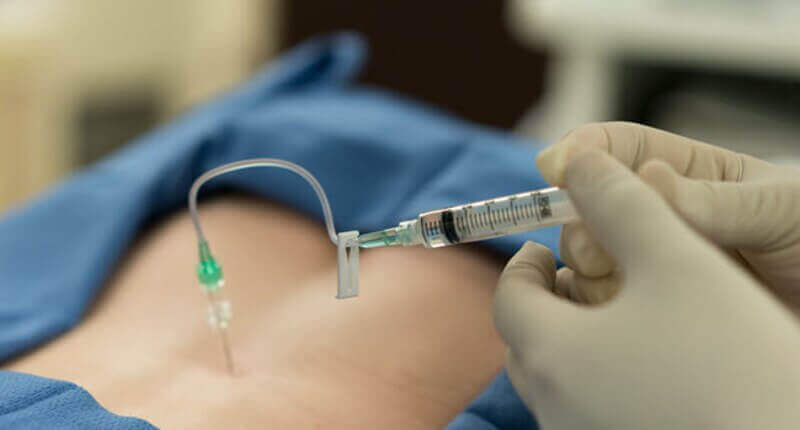
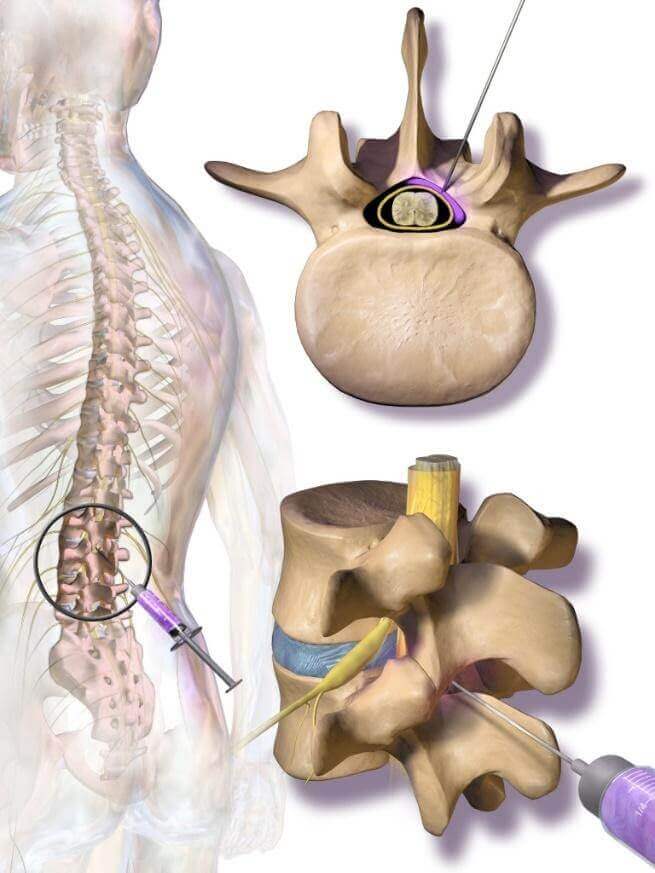
Injection under image guidance. We inject under fluoroscopic or ultrasound guidance. For intra-articular work — hip, knee, shoulder — fluoroscopy confirms needle placement in the joint space. For tendons and ligaments, dynamic ultrasound allows us to see the needle tip in real time and place the material exactly where the tissue damage is. Accuracy here is non-negotiable. Blind injections of regenerative biologics are, in my view, not acceptable clinical practice.

Recovery. The harvest site has a small dressing and is mildly sore for 2–3 days. The injected area may ache for 1–2 weeks as the biological response begins. We advise protected weight-bearing for joint procedures and physiotherapy commencing at 2–3 weeks.

Nanofat in Avascular Necrosis — A Personal Perspective

I want to spend some time here because AVN is a condition that sits very close to my clinical heart. We see a significant number of AVN patients at IBAP Clinics, and what strikes me repeatedly is how late they are referred. By the time many patients arrive — having been told "only joint replacement will fix this" — we are already managing Ficat Stage III or IV, where the femoral head has collapsed. At that stage, our options are genuinely limited.

But for Stage I and Stage II AVN, the biology of regenerative medicine is a rational, scientifically supported intervention. The primary pathology in AVN is vascular — ischaemia leading to osteonecrosis. If we can restore blood supply, stimulate osteoprogenitor activity, and reduce the inflammatory cycle that accelerates collapse, we change the natural history. That is not a marketing claim. That is the mechanistic logic underpinning the combination of:

  • Nanofat (pericytes for angiogenesis, MSCs for osteoprogenitor support)
  • BMAC — bone marrow aspirate concentrate (direct osteogenic progenitors)
  • PRP (growth factor scaffolding)
  • Pharmacological therapy — bisphosphonates, teriparatide, statins and prostacyclin analogues in appropriate patients

The pharmacological component is, frankly, underemphasised in most regenerative medicine discussions. At IBAP Clinics, our outcomes in AVN have been meaningfully better, I believe, because we treat the whole biology — not just inject and hope. We have had patients come to us scheduled for total hip replacement within three months, who, at twelve months following our biological cocktail protocol, have returned to near-normal function with preserved femoral head architecture on MRI.

🌟 Analogy
AVN of the femoral head is like a tree whose roots are being slowly strangled by drought. Injecting stem cells alone is like giving the tree water once — helpful, but insufficient if the underlying drought is not addressed. Our combination approach — regenerative cells plus pharmacological therapy to improve the soil conditions — is the difference between survival and thriving.

Clinical Indications — Evidence and Strength of Support

ConditionEvidence LevelTypical ImprovementCombine With
Avascular Necrosis (hip, knee, shoulder) — Stage I–IILevel II–III (prospective series)60–80% functional improvementBMAC + PRP + pharmacological
Osteoarthritis (knee, hip, shoulder) — early/moderateLevel I–II (RCTs, meta-analyses)Significant pain reduction; functional gainPRP ± HA
Rotator Cuff Tendinopathy / Partial TearLevel II–IIIGood in partial tears; modest in full-thicknessPRP + physiotherapy
Lateral Epicondylitis / Achilles TendinopathyLevel IIComparable or superior to corticosteroidPRP
Plantar Fasciitis (recalcitrant)Level II–IIIModerate; useful after PRP non-responsePRP ± shockwave
Ligament Laxity / Partial TearsLevel IIIModerate; adjunct to rehabilitationPhysiotherapy
Muscle Tears / Intramuscular FibrosisLevel IIIEmerging evidence; promising in chronic tearsPhysiotherapy + PRP
Discogenic Low Back PainLevel III (pilot studies)Early positive signals; not standard of careIntradiscal PRP

Nanofat vs Other Regenerative Options — A Practical Comparison

FeatureNanofatPRPBMACExosomes
SourceAdipose tissue (fat)Peripheral bloodBone marrow (iliac crest)Donor/allogenic (India: grey area)
Contains living MSCsYes — MSCs, pericytesNoYes — osteogenic MSCsNo (cell-derived vesicles)
Growth factorsRich — VEGF, HGF, IGF-1High — PDGF, TGF-β, EGFModerateConcentrated signalling molecules
Harvest discomfortModerate (local anaesthetic)Minimal (venepuncture)Moderate-high (bone marrow)None (pre-prepared)
Volume of materialModerate (3–10 ml injectable)3–8 ml5–10 ml concentrate1–2 ml vial
Legal/regulatory IndiaSame-day autologous: permissibleEstablished, permissibleSame-day autologous: permissibleNot yet regulated; exercise caution
Best combined withPRP, BMAC, pharmacologicalNanofat, HA, BMACNanofat, PRPResearch setting only (India)

Legality in India — What You Need to Know

This is a topic I feel strongly about, because confusion in this space has harmed patients. Let me be clear about what is and is not permissible under current Indian regulation.

The CDSCO (Central Drugs Standard Control Organisation) and ICMR (Indian Council of Medical Research) have jointly issued guidelines (2019, updated 2022) governing stem cell research and therapy. The key distinction is between minimally manipulated and more-than-minimally-manipulated cells.

Nanofat — as we prepare it at IBAP Clinics — falls within the permissible category when it is: (1) autologous (from the same patient), (2) prepared and re-administered in the same surgical procedure, (3) subject to mechanical processing only (no chemical reagents, no in-vitro culture, no cryopreservation for later use), and (4) used in a homologous manner (fat-derived cells into musculoskeletal tissue — a biologically related target).

What is not permissible without approved clinical trial registration (CTRI) is: culture-expanded adipose MSCs, allogeneic (donor) stem cells, or the use of chemical agents to expand or isolate cell populations outside a licensed facility. I mention this because I have seen clinics in India offering "cultured stem cell therapy" or "exosome therapy from donor sources" without any CTRI registration — which is both legally questionable and, frankly, ethically concerning.

At IBAP Clinics, we operate within the regulatory framework. We do not offer therapies that have not cleared the appropriate legal and ethical thresholds.

What Results Can You Realistically Expect?

I will give you an honest answer rather than a promotional one, because I think patients deserve that.

Nanofat is not a magic cure. It is a powerful adjunct to a comprehensive treatment plan. For patients with early-to-moderate joint degeneration, tendinopathy, or ligament injury who have not responded adequately to conventional therapy, the realistic expectation is:

  • 3–6 months for the biological effect to peak — regenerative therapies work slowly, through cellular differentiation and tissue remodelling, not through pharmacological blocking of pain signals
  • Meaningful pain reduction (VAS reduction of 30–50%) in most responders at 6 months
  • Functional improvement — better range of motion, return to physical activity — in the majority
  • Imaging changes — cartilage preservation, tendon structural improvement — in a proportion; not universal
  • Durability — most studies show effects lasting 12–24 months; repeat procedures are an option

For AVN in particular, our combination protocol has produced outcomes I find genuinely encouraging — particularly in patients who commit to the full pharmacological protocol alongside the biological injections. I want to be cautious about citing individual case outcomes, because good science demands population-level data, and we are working towards publishing our series. What I can say is that early-stage AVN, treated comprehensively, can have its natural history materially altered.

💬 Dr Vijay's View
I believe the single biggest predictor of poor outcomes in regenerative medicine is not the treatment — it is the timing. Patients who arrive at Stage III AVN or grade IV OA are asking biology to do something structural surgery does better. The role of every GP and orthopaedic colleague is early referral. The role of every patient is not dismissing pain as "just wear and tear."

Frequently Asked Questions

What is Nanofat therapy?
Nanofat is an ultra-fine emulsion of your own adipose (fat) tissue, created by mechanically filtering microfat through fine sieves. It concentrates stromal vascular fraction (SVF) cells — including mesenchymal stem cells and pericytes — for injection into damaged joints, tendons, ligaments or skin. No chemicals are used; no laboratory processing outside the clinic theatre.
How is Nanofat different from PRP?
PRP is derived from blood and delivers growth factors and platelets — important signalling molecules but no living stem cells. Nanofat is derived from fat and delivers living MSCs, pericytes and growth factors simultaneously. They are complementary, and at IBAP Clinics we often use them together for a more comprehensive biological response.
Is Nanofat therapy legal in India?
Same-day autologous adipose tissue, mechanically processed and re-injected within the same surgical procedure for a homologous indication, is permissible under current CDSCO and ICMR guidelines. Culture-expanded cells, allogeneic (donor) cells, and chemical manipulation without CTRI registration are not. IBAP Clinics operates fully within the current regulatory framework.
Does the procedure hurt?
The harvest is performed under local anaesthesia (tumescent technique) from the abdomen or inner thigh. The infiltration of local anaesthetic is the most uncomfortable part — roughly comparable to a dental injection. The actual fat harvest and subsequent injection are usually well tolerated. Post-procedure soreness at the harvest site typically resolves within 2–3 days.
Who is a good candidate for Nanofat?
Patients with early-to-moderate AVN, osteoarthritis, rotator cuff tendinopathy, ligament laxity, plantar fasciitis, muscle tears, or chronic tendon pain who have not responded sufficiently to conventional therapy are the best candidates. Advanced structural destruction (collapsed femoral head, bone-on-bone end-stage OA) reduces the scope for biological therapy — which is why early referral matters enormously.
How many sessions are needed?
The majority of patients at IBAP Clinics receive a single procedure and are reviewed at 3 months and 6 months. A second session may be offered at 3–6 months for moderate-to-severe conditions or partial responders. Results continue to evolve for up to 6 months after treatment, so patience is essential — do not judge the outcome at 6 weeks.
Can Nanofat be combined with other regenerative treatments?
Yes — and at IBAP Clinics, combination is often the standard for complex or severe cases. Nanofat is frequently combined with PRP, hyaluronic acid, or BMAC (bone marrow aspirate concentrate). For AVN, we additionally employ a pharmacological protocol including bisphosphonates, statins and, in selected patients, teriparatide or prostacyclin analogues. This comprehensive approach is, we believe, the reason our AVN outcomes compare favourably with published series.

Ready to Explore Nanofat Therapy?

Book a regenerative medicine consultation at IBAP Clinics, Hyderabad. Dr Vijay and the team will review your imaging, explain your options honestly, and design a protocol appropriate for your condition and stage.

IBAP Clinics Main
2nd Floor, 284/A, Road No. 12
Above IDFC First Bank, near Omega Hospitals
MLA Colony, Banjara Hills
Hyderabad 500034
IBAP Clinics Branch
Sy No. 2, 4th Floor, Plot No. 200
Beside South India Shopping Mall
Opp. Fortune Heights, Mythri Nagar
Madeenaguda, Hyderabad 500049

References

  1. Gimble JM, Katz AJ, Bunnell BA. Adipose-derived stem cells for regenerative medicine. Circ Res. 2007;100(9):1249–1260.
  2. Crisan M, Yap S, Casteilla L, et al. A perivascular origin for mesenchymal stem cells in multiple human organs. Cell Stem Cell. 2008;3(3):301–313.
  3. Tonnard P, Verpaele A, Peeters G, et al. Nanofat grafting: basic research and clinical applications. Plast Reconstr Surg. 2013;132(4):1017–1026.
  4. Caplan AI, Correa D. The MSC: an injury drugstore. Cell Stem Cell. 2011;9(1):11–15.
  5. Bora P, Majumdar AS. Adipose tissue-derived stromal vascular fraction in regenerative medicine: a brief review on biology and translation. Stem Cell Res Ther. 2017;8(1):145.
  6. Kim M, Kim I, Lee SK, Bang SI, Lim SY. Clinical trial of autologous differentiated adipocytes from stem cell-derived preadipocytes in treating avascular necrosis of the femoral head. Dermatol Surg. 2014;40(1):33–38.
  7. Pak J, Lee JH, Pak HS, et al. Regenerative effects of stromal vascular fraction cells in treatment of osteoarthritis. J Stem Cell Res Ther. 2014.
  8. Usuelli FG, D'Ambrosi R, Maccario C, et al. Adipose-derived stem cells in orthopaedic pathologies. Br Med Bull. 2017;124(1):31–54.
  9. ICMR/DBT Guidelines for Stem Cell Research. Indian Council of Medical Research / Department of Biotechnology, India. 2017; updated 2022.
  10. Lamo-Espinosa JM, Mora G, Blanco JF, et al. Intra-articular injection of two different doses of autologous bone marrow mesenchymal stem cells versus hyaluronic acid in the treatment of knee osteoarthritis. J Transl Med. 2016;14(1):246.
  11. Hauser RA, Orlofsky A. Regenerative injection therapy with whole bone marrow aspirate for degenerative joint disease. Clin Med Insights Arthritis Musculoskelet Disord. 2013;6:65–76.
  12. Péault B, Zuk P, Erickson HP, et al. Stem and progenitor cells in skeletal muscle development, maintenance, and therapy. Mol Ther. 2007;15(5):867–877.
⚖️ Medical Disclaimer
This article is written for educational purposes by Dr Vijay Bhaskar Bandikatla and does not constitute personalised medical advice. Nanofat and other regenerative therapies should only be considered following a full clinical assessment by a qualified specialist. Results vary between individuals depending on diagnosis, stage of disease, and overall health. IBAP Clinics operates within current ICMR and CDSCO regulatory guidelines. Patients are encouraged to ask questions and obtain informed consent documentation before any procedure. If you are in pain, please seek professional evaluation — do not self-diagnose or self-treat.

Happy Patients - Live Testimonials

Meenakshi, 75 years - Relived From Knew Pain

Mrs. Naga Sathyavathi relieved from Back Pain & Shoulder Pain

Mr. Bhaskar Reddy inspiring recovery from L1 Spine Issue.

Raghav Reddy recovered from Pain to Relief

Mr. Chakrapani recovered from Severe Joint and Neck Pain

Happy Patients - Google Reviews

Dr. Vijay Bhaskar Bandikatla

Founder IBAP Clinics, Pain Physician MBBS · DA · FRCA (London) · FFPMRCA (Pain Medicine) · CCT (UK) · Advanced Pain Training (Cambridge) · Fellowship in Neuromodulation & Advanced Pain (London) · DDSMed (Sports Medicine, Pune ISST— ISPA, Chicago) MBA (Hospital Management)

Dr Vijay Bhaskar Bandikatla

Founder & Interventional Pain Specialist — IBAP Clinics, Hyderabad
MBBS · DA · FRCA (London) · FFPMRCA (Pain Medicine, UK) · MBA (Hospital Management)
CCT (Anaesthesia & Pain Medicine, UK) · Advanced Pain Training (Cambridge University Hospitals)
DDSMed Sports Medicine (Chicago) · Fellowship in Neuromodulation & Advanced Pain (London)

Dr Vijay brings over 15 years of postgraduate training across the United Kingdom’s most prestigious institutions — including the Royal College of Anaesthetists, Cambridge University Hospitals, and a dedicated neuromodulation fellowship in London — to his practice in Hyderabad. He is one of very few clinicians in India trained to the level of FFPMRCA — the Faculty of Pain Medicine of the Royal College of Anaesthetists — the highest qualification in pain medicine available in the UK.

His specialist expertise spans the full spectrum of knee pain management: from precision PRP and BMAC injections to cooled radiofrequency genicular nerve ablation, intrathecal drug delivery, and spinal cord stimulation for refractory pain states. He manages cases ranging from the weekend cricketer’s torn meniscus to the elderly cardiac patient with end-stage OA who has been told there are no further options.

Epidural Spinal Injections

Epidural Spinal Injections

Epidural injections involve the injection of medication, usually a combination of a local anesthetic and a corticosteroid, into the epidural space around the spinal cord. This procedure is commonly used to alleviate pain and inflammation associated with conditions such as herniated discs, spinal stenosis, and sciatica. The local anaesthetic provides immediate pain relief by numbing nerves, while the corticosteroid helps reduce inflammation for longer-term effects. The epidural space is the outermost part of the spinal canal, located just outside the protective membrane called the dura mater.The injection is typically administered by a qualified healthcare professional, such as an anesthesiologist or pain management specialist. The goal of an epidural spinal injection is to reduce inflammation and alleviate pain caused by various conditions affecting the spine and surrounding tissues
Close-up of the injection site for epidural spinal injections

Some common reasons for undergoing this procedure include:

  • Herniated Disc: When the soft inner material of a spinal disc protrudes through the tough outer layer, it can irritate nearby nerves, causing pain.
  • Spinal Stenosis: This is a narrowing of the spinal canal, which can put pressure on the spinal cord and nerves, leading to pain and discomfort.
  • Degenerative Disc Disease: As the discs between the vertebrae age and break down, they can contribute to pain and inflammation.
  • Sciatica: Inflammation or compression of the sciatic nerve, which runs from the lower back down the back of each leg, can cause pain, numbness, and tingling.
  • Spinal Arthritis: Inflammatory conditions affecting the spine, such as ankylosing spondylitis or osteoarthritis, can lead to pain and stiffness.

Our goal is to help the patient regain their quality of life

In our pain clinic, we provide pain relief so you can regain your identity.

Chat with us

Book An Appointment

Choose Branch