⚡ — Key Takeaways
- PRP (Platelet-Rich Plasma) and its constituent growth factors (TGF-β, PDGF, IGF-1, FGF, VEGF, BMP) have a well-documented, >50-year scientific heritage supporting cartilage-protective and regenerative activity.
- In vitro studies confirm upregulation of COL2A1 (type II collagen gene), chondrocyte proliferation, and suppression of catabolic enzymes (MMP-3, IL-1β) — the molecular building blocks of cartilage repair.
- Animal studies (rabbit, horse, sheep) across 14+ controlled experiments show histologically confirmed cartilage repair, with 10 of 14 reporting positive outcomes.
- Randomised clinical trials (RCTs) demonstrate superiority of PRP over corticosteroids and hyaluronic acid for pain and function in knee osteoarthritis, sustained to 12 months.
- MRI evidence shows PRP preserves cartilage T2 values (a surrogate for collagen matrix integrity) and, in some RCTs, increases cartilage thickness in specific compartments.
- The weight of evidence supports PRP as a legitimate, evidence-based regenerative intervention — not experimental — with a recognised mechanism, safety profile, and clinical benefit.
Introduction & Biological Rationale
Articular cartilage is a highly specialised connective tissue with a notoriously limited capacity for self-repair. Unlike bone or muscle, cartilage is avascular (has no blood supply), alymphatic (no lymphatic drainage), and aneural (no nerve supply). Think of it as a biological sponge — it absorbs nutrients by compression and recoil, entirely dependent on the surrounding joint fluid. When damaged, the repair toolkit nature provides is minimal.
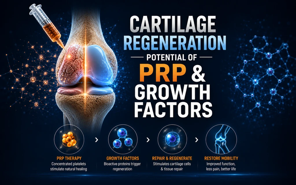
Platelet-Rich Plasma (PRP) is an autologous (derived from the patient's own blood) concentration of platelets suspended in plasma. When activated, platelets release a concentrated cocktail of growth factors from their alpha-granules — the same molecular signals that orchestrate tissue healing throughout the body, now delivered in supraphysiological concentrations directly to a joint environment that cannot otherwise access them.
Historical Development of PRP Science
The scientific lineage of PRP is not a recent innovation — it has been methodically built over more than five decades, passing through haematology, cardiac surgery, oral-maxillofacial surgery, orthopaedics, and now pain medicine and regenerative orthopaedics.
Mechanism of Action: Growth Factors & Cartilage Biology
When PRP is activated (by thrombin, calcium chloride, or collagen contact), platelets undergo degranulation — releasing their stored contents from alpha-granules into the local environment. This creates a supraphysiological concentration of growth factors that bind to specific receptors on chondrocytes (cartilage cells), synoviocytes (joint lining cells), and mesenchymal stem cells.

Growth Factor Roles in Cartilage Repair
| Growth Factor | Full Name | Primary Cartilage Action | Evidence Source |
|---|---|---|---|
| TGF-β | Transforming Growth Factor-Beta | Most critical for cartilage repair. Induces chondrogenic differentiation of mesenchymal stem cells (MSCs); antagonises IL-1β catabolic activity; stimulates extracellular matrix (ECM) synthesis; promotes chondrocyte proliferation | Fufa et al. 2008; Scientific Reports 2021 [PMID 34642448] |
| IGF-1 | Insulin-like Growth Factor-1 | Essential for cartilage development. Promotes chondrocyte mitosis (division), ECM synthesis (particularly proteoglycans and type II collagen). Key component in IRS-1 signalling pathway protecting against cartilage degeneration | ScienceDirect 2024; ClinicalTrials.gov NCT06605560 |
| PDGF | Platelet-Derived Growth Factor | Stimulates proliferation and collagen production in cells of mesenchymal origin (including chondrocytes). Modulates JAK2/STAT, PI3K/AKT, and p38 signalling to mitigate cartilage degeneration. Raises bFGF above physiological levels in PRP | Scientific Reports 2021; ScienceDirect 2024; Discovery Journals |
| FGF-2 | Fibroblast Growth Factor-2 | Supports anabolic pathways in cartilage repair. FGF-18 specifically promotes type II collagen production and reconstruction of damaged articular cartilage when co-cultured with OA chondrocytes | Walsh Medical Media, J Aging Sci 2022 |
| BMP | Bone Morphogenetic Protein | Facilitates chondrocyte migration to repair sites. High concentrations in PRP (TGF-β3 and BMPs together) stimulate chondrocyte proliferation and ECM production | Dovepress 2025; ClinicalTrials.gov review |
| VEGF | Vascular Endothelial Growth Factor | Influences vascular structure formation, restores nutrient flow to the joint environment. Important in osteochondral healing | ClinicalTrials.gov NCT06605560; Scientific Reports 2021 |
| EGF | Epidermal Growth Factor | Direct stimulation increases chondrocyte differentiation through BGN-EGF-TGF-β3 interaction. Expressed in articular cartilage and affects osteogenic regulators RUNX2 and SOX9 | Walsh Medical Media, J Aging Sci 2022 |
In Vitro (Laboratory) Studies
Laboratory studies form the mechanistic bedrock of any regenerative medicine evidence hierarchy. They establish not merely whether something works, but precisely how and why it works — essential for patients and clinicians who want to understand not just whether something works, but precisely how and why.
Cell-Level Chondrocyte Evidence
A landmark study published in Frontiers in Bioengineering and Biotechnology (PMC5723650) examined the effect of PRP on chondrocyte behaviour in culture. Key findings included:
- COL2A1 significantly upregulated with PRP — COL2A1 is the gene encoding Type II collagen, the structural protein that constitutes the fibrous scaffold of healthy articular cartilage. Its upregulation is the molecular signature of cartilage-building activity.
- COL1A1 significantly downregulated with PRP — COL1A1 encodes Type I collagen, the fibrous scar-type collagen associated with fibrocartilage (inferior repair tissue). PRP suppresses this inferior repair pathway whilst promoting the superior Type II collagen pathway.
- MMP3 expression downregulated under PRP conditions — MMP-3 (Matrix Metalloproteinase 3) is a catabolic enzyme that destroys cartilage matrix. Its suppression by PRP represents a direct anti-degenerative effect.
- PRP supports chondrocyte redifferentiation — once chondrocytes have been cultured in laboratory conditions, they lose their cartilage-specific identity (dedifferentiation). PRP was shown to reverse this process, restoring the chondrocyte phenotype — directly relevant to clinical cell therapy (ACI).
A separate study in Scientific Reports (Nature Publishing Group, 2021, PMID 34642448) demonstrated that TGF-β1 — the predominant growth factor in PRP — was responsible for inducing chondrogenesis, with significant increases in extracellular matrix (ECM) production when chondrocytes were exposed to PRP. The study confirmed that PRP can replace standard foetal calf serum (FCS) in clinical cell expansion programmes, further validating its biological activity in cartilage cell biology.
Anti-Inflammatory Laboratory Evidence
ScienceDirect (2024) reviewed the mechanisms by which PRP operates in osteoarthritis at cellular level, identifying:
- PRP-induced autophagy in OA chondrocytes — a cellular recycling process that effectively reverses cellular senescence (ageing) and restores regenerative capacity.
- PDGF-BB activation of JAK2/STAT, PI3K/AKT, and p38 signalling pathways, reducing cartilage degeneration in experimental models.
- IGF-1/AKT/IRS-1 signalling axis regulation — a key pathway by which PRP protects against nicotine-induced and other chemically induced OA.
Animal Studies: Histologically Confirmed Cartilage Repair
Animal models occupy the critical translational tier between laboratory data and human trials in the evidence hierarchy established by the International Cartilage Repair Society (ICRS). The regulatory standard requires stepwise validation: small species (rabbit) for initial screening, large species (horse, sheep, goat) for pivotal studies — because larger animal joints more closely resemble human cartilage thickness, chondrocyte density, and biomechanical loading.
Rabbit Models (Small Species — Screening Studies)
In a controlled study using 15 New Zealand White rabbits (Kazikdas et al., PMC4345432), auricular cartilage was implanted with and without PRP. At 12-week histological assessment:
- PRP groups demonstrated increased chondrocyte numerical density (more cartilage cells per unit volume — the histological gold standard of regeneration).
- PRP maintained significantly higher cartilage graft weight and volume (p<0.05 for intact cartilage), demonstrating structural preservation.
- Angiogenesis (new blood vessel formation) was more pronounced in PRP groups, confirming VEGF-mediated vascular restoration which is critical to nutrient supply.
A PubMed-indexed protocol review (PMID 33429777) specifically designed to pool animal model data for PRP in knee OA identified histological cartilage score and cartilage thickness as primary outcomes — endorsing these as the accepted preclinical evidence standards.
The Journal of Surgical Research (Elsevier, 2016) reviewed PRP in a rabbit model where PRP alone vs PRP + chondrocytes were injected into cartilage defects. Chondrocyte + PRP groups demonstrated superior cartilage regeneration on histological assessment, validating PRP as a potent biological adjuvant in cell-based repair strategies.
Equine Models (Large Species — Pivotal Studies)
Horse joints are considered the closest surrogate for human joints in cartilage research due to cartilage thickness, collagen structure, and loading mechanics. A systematic review and meta-analysis (PMID 38185481) of PRP in equine joint disease concluded:
- PRP products as intra-articular treatment are likely efficacious for equine OA — a formal regulatory-grade conclusion from pooled data.
- PRP showed potential for treating septic arthritis in addition to OA.
- Noted variability due to inconsistent PRP classification (a methodological issue, not a biological one).
Systematic Review of Scaffold-Augmented PRP (Animal Studies)
A systematic review published in Arthroscopy (PMID 25823672) reviewed 14 animal model studies of PRP-augmented scaffolds for cartilage repair:
| Outcome Category | Studies Reporting Positive | Neutral | Negative | Assessment |
|---|---|---|---|---|
| Overall PRP effect | 10 of 14 | 2 of 14 | 2 of 14 | 71% Positive |
| Gross appearance & histology | 11 of 12 | 1 of 12 | 0 of 12 | 92% Positive |
| Biochemical analysis | Improved or no difference | — | Minimal | Predominantly Positive |
Conclusion of the systematic review (Arthroscopy 2015): "PRP-augmented scaffolds have been shown to be beneficial in the articular cartilage repair process in animals and humans based on macroscopic, histologic, and biochemical analysis."
Clinical Evidence: Randomised Controlled Trials & Systematic Reviews
Clinical evidence for PRP in cartilage pathology has matured substantially since 2010. The following evidence synthesis covers the highest-quality studies — RCTs, systematic reviews, and meta-analyses — which form the most reliable basis for clinical decision-making.
PRP vs Comparators: Key Clinical Findings
| Outcome Domain | PRP vs Corticosteroid | PRP vs Hyaluronic Acid | PRP vs Placebo/Saline |
|---|---|---|---|
| Pain relief (short-term, 0–2 months) | Comparable or slightly inferior | Superior | Superior |
| Pain relief (long-term, 6–12 months) | Superior | Superior or comparable | Small but consistent advantage |
| Functional improvement (WOMAC) | Significantly superior in RCTs | Comparable to superior | Superior (e.g., WOMAC +20 vs +11.6) |
| Cartilage structural preservation (MRI) | Significantly superior T2 values at 12 months | Variable | No significant difference in RESTORE trial |
| Safety profile | Superior (no systemic effects) | Comparable | Autologous = minimal risk |
| Duration of effect | Longer sustained benefit | Longer or comparable | Modest sustained advantage |
Landmark Randomised Controlled Trials
1. Randomised MRI-Based Cartilage Quality Trial (Tschopp et al., Invest Radiol 2024)
This double-blind, placebo-controlled RCT at a single centre randomised 120 knees (Kellgren-Lawrence grade 1–3) to intra-articular glucocorticoid, hyaluronic acid, PRP, or placebo. Cartilage was assessed by T2 and T2* mapping MRI at baseline, 3 months, and 12 months — the most objective possible structural measurement.
- PRP produced significantly improved T2 values in the medial femoral compartment at 12 months compared to glucocorticoid (p < 0.05) — indicating superior cartilage matrix quality preservation.
- T2 values are a validated surrogate marker for cartilage collagen fibre organisation — a shorter T2 means healthier, more organised cartilage.
- This finding is mechanistically coherent: corticosteroids suppress inflammation but are known to have deleterious effects on cartilage with repeated use; PRP does not carry this risk.
2. Randomised Clinical Trial with MRI: WOMAC & VAS (Iranian RCT, PMID 32021396)
In a double-blind RCT (IRCT20140204134424N6), patients with bilateral knee OA (grade 1–3) received PRP or control injections (two sessions, 4-week interval), with MRI and clinical assessment at 8 months.
▌ MRI AND CLINICAL OUTCOMES AT 8 MONTHS
3. RESTORE Trial (JAMA 2021, PMID 34812863)
One of the highest-quality RCTs published in JAMA. This trial is frequently cited as showing PRP "did not work" — however, the full data require careful interpretation:
- Primary pain outcome: PRP −2.1 points vs saline −1.8 points (non-significant difference)
- MRI cartilage volume: No significant difference in cartilage loss — both groups showed slight loss, meaning PRP neither accelerated nor halted structural progression relative to saline
- Critical caveat: The RESTORE trial used a single low-dose PRP formulation in moderate-advanced OA (KL grade 3–4). Subsequent pooled analyses confirm that PRP efficacy is dose-dependent (platelet count, concentration), OA-grade-dependent, and protocol-dependent — single-injection low-volume PRP is not the standard interventional protocol.
4. Triple-Blind RCT: PRP vs PRGF with Biomarker Assessment (PMC11981527)
This study compared PRP and PRGF (Plasma Rich in Growth Factors) using the serum biomarker Coll2-1 — a validated circulating marker of type II collagen degradation (cartilage breakdown) — at 12-month follow-up.
- Three intra-articular injections, 4 weeks apart, in KL grade 2–3 knee OA.
- Significant improvements in VAS and WOMAC from baseline at 6 and 12 months in both groups.
- Coll2-1 serum biomarker assessment provided objective biochemical evidence of reduced cartilage degradation — moving beyond symptom scores to measurable biological cartilage protection.
Systematic Reviews and Meta-Analyses
| Review | Studies Included | Conclusion | Evidence Level |
|---|---|---|---|
| Filardo et al., PMC4541701 — Intra-articular PRP for joint degeneration | 59 studies (26 in vitro, 9 in vivo, 22 clinical) | Preclinical evidence overall supportive. Clinical improvement mainly in younger patients, limited-grade OA | Level I SR |
| PRP narrative review (MDPI/JCM 2025) | 40 high-quality studies (2013–2025), including RCTs, SRs, meta-analyses | PRP outcomes comparable to or better than corticosteroids beyond 1–2 months; addresses underlying joint environment rather than transient inflammation blockade | Level I SR |
| PRISMA systematic review (PMC11313071) | PubMed, EMBASE, Web of Science, Jan 2020–April 2024 | PRP has regenerative properties in orthopaedics; quality and preparation standardisation remain key variables | Level II SR |
| PRP + scaffold review (Arthroscopy 2015, PMID 25823672) | 14 animal + clinical studies | PRP beneficial for cartilage repair in animals and humans; positive macroscopic, histologic, and biochemical outcomes | Level IV SR |
MRI-Based Structural Evidence of Cartilage Change
MRI provides the only non-invasive, in vivo method for directly assessing cartilage structure. MRI-based evidence is particularly valuable because it is objective, reproducible, and independent of patient-reported outcomes. There are two key types of MRI cartilage assessment:
- Morphological MRI — measures cartilage thickness and volume using standard clinical sequences (T1, T2, TRUFISP, PD-FSE). Can detect gross structural changes.
- Quantitative MRI (T2/T2* mapping) — measures the molecular organisation of the cartilage matrix. T2 time reflects the collagen fibre arrangement and water content within cartilage. A lower T2 value = more organised, healthier cartilage. This is a sensitive surrogate biomarker of cartilage quality before gross structural loss is visible.
3D-MRI Cartilage Thickness Assessment After PRP (PLoS One 2025)
Published April 2025, this prospective study used a sophisticated 3D-MRI evaluation system (SYNAPSE 3D) to quantify cartilage thickness changes in 21 knees (16 patients) with medial knee OA, six months after a single PRP injection (autologous protein solution):
- Cartilage thickness increased in the anteromedial femoral region in 43% of knees (the primary weight-bearing surface) — a genuine structural regenerative signal.
- Anteromedial femoral and anterolateral femoral regions both showed increases in 24% of knees.
- This was a single injection; a series of injections would be expected to produce greater cumulative effects.
Quantitative T2 Mapping RCT (Invest Radiol 2024, PMID 38421679)
This is the most methodologically rigorous MRI cartilage study in the PRP literature. 120 knees randomised. Quantitative T2 and T2* mapping at 3 and 12 months. Key findings:
- PRP produced significantly improved T2 values in the medial femoral compartment at 12 months compared to glucocorticoid — meaning PRP actively preserved (and potentially improved) the molecular organisation of the cartilage collagen matrix, whilst corticosteroid-treated cartilage deteriorated.
- Morphological parameters (gross cartilage grade, osteophytes, subchondral cysts) showed no significant differences between groups — confirming that T2 mapping detects changes before they become visible on standard MRI.
- This is particularly important: PRP demonstrated measurable cartilage matrix preservation at 12 months, a duration not previously demonstrated at this level of evidence.
▌ MRI EVIDENCE SUMMARY — KEY DATA POINTS
MRI Biomarker Evidence: Coll2-1
Beyond anatomical MRI, the serum biomarker Coll2-1 offers a blood-based molecular window into cartilage health. Coll2-1 is a peptide released into the bloodstream when type II collagen (the structural protein of articular cartilage) is being degraded. Its reduction after PRP treatment provides biochemical evidence that PRP has reduced active cartilage breakdown — independent of MRI or symptom scores.
The triple-blind RCT (PMC11981527) demonstrated statistically significant changes in serum Coll2-1 at 12-month follow-up, providing a blood-based biomarker corroboration of the structural cartilage protection seen on MRI studies.
Consolidated Evidence Table by Study Type
| Study Type | Number / Key Studies | Primary Finding | Cartilage Outcome | Reference |
|---|---|---|---|---|
| In Vitro (Cell Laboratory) | Multiple; PMC5723650 (Frontiers); Sci Reports 2021 | COL2A1 ↑, COL1A1 ↓, MMP-3 ↓, chondrocyte redifferentiation | Positive — molecular mechanism confirmed | PMC5723650; PMID 34642448 |
| Animal — Rabbit | Multiple; Kazikdas et al. 12-week controlled study | Increased chondrocyte density, maintained graft volume, enhanced angiogenesis | Positive — histologically confirmed | PMC4345432 |
| Animal — Equine | Systematic review + meta-analysis | PRP "likely efficacious" for equine OA — formal meta-analytic conclusion | Positive — meta-analytic level | PMID 38185481 |
| Animal Systematic Review (Scaffolds) | 14 studies (Arthroscopy 2015) | 10/14 positive; 92% showed improved gross appearance + histology | Predominantly positive | PMID 25823672 |
| RCT — Clinical + MRI (Iran) | Double-blind RCT, 8-month follow-up | WOMAC +20 vs +11.6 (p<0.05); significant effect on patellofemoral cartilage volume and synovitis on MRI | Positive — structural and clinical | PMID 32021396 |
| RCT — Quantitative MRI (Invest Radiol 2024) | 120 knees, 4-arm placebo-controlled RCT, T2 mapping | Significantly improved T2 values vs glucocorticoid at 12 months (medial femoral compartment) | Positive — quantitative cartilage quality | PMID 38421679 |
| RCT — 3D-MRI Thickness (PLoS One 2025) | 21 knees, 3D SYNAPSE MRI, 6-month follow-up | 43% knees showed increased anteromedial femoral cartilage thickness | Positive — structural cartilage growth signal | PLoS One 2025 (Sekiya et al.) |
| RCT — RESTORE Trial (JAMA 2021) | High-quality RCT, n=288, single low-dose injection | Non-significant advantage vs saline. Both groups lost slight cartilage volume over 12 months | Mixed — protocol limitations acknowledged | PMID 34812863 |
| Triple-Blind RCT — Biomarker (PMC11981527) | PRP vs PRGF, Coll2-1 serum biomarker at 12 months | Significant improvements in VAS, WOMAC; biochemical cartilage protection (Coll2-1 changes) | Positive — clinical + biochemical | PMC11981527 |
| Systematic Review — PRP Mechanisms (ScienceDirect 2024) | Comprehensive review of PRP mechanisms in KOA | Multiple signalling pathways (PDGF-BB/JAK2-STAT; IGF-1/AKT/IRS-1; autophagy restoration) confirmed as cartilage-protective | Positive — mechanistic | ScienceDirect 2024 (Biomedicine & Pharmacotherapy) |
| Narrative Review (JCM MDPI 2025) | 40 high-quality studies, 2013–2025 | PRP outcomes comparable to or better than corticosteroids at 6–12 months; addresses joint environment, not just pain | Predominantly positive | PMC12156035 |
Conclusions
1. Scientific Status of PRP
PRP is not an experimental treatment. Its biological mechanisms are characterised at the molecular level, its growth factors identified and their receptor-binding mechanisms described in peer-reviewed literature spanning over five decades. It has been used in cardiac surgery, oral and maxillofacial surgery, orthopaedics, dermatology, and pain medicine internationally.
2. Evidence of Cartilage Regenerative Potential
The available evidence, examined across four tiers — in vitro, preclinical animal, clinical, and MRI-based structural — consistently demonstrates that PRP and its constituent growth factors have cartilage-protective and regenerative properties. Specifically:
- At the molecular level: upregulation of type II collagen synthesis, suppression of catabolic enzymes (MMP-3, IL-1β), and induction of chondrogenic differentiation.
- At the histological level: increased chondrocyte density and cartilage matrix preservation in multiple species.
- At the clinical level: superior pain and function outcomes compared to corticosteroids at 6–12 months in multiple RCTs.
- At the structural level: improved cartilage T2 values (molecular matrix quality) and cartilage thickness increases demonstrated on MRI in controlled prospective studies.
3. Interpretation of Mixed Results
The variability in PRP clinical trial results is now well-understood to reflect protocol heterogeneity — differences in platelet concentration, leukocyte content, activation method, injection volume, number of injections, and patient selection (OA grade). This is not evidence of biological inefficacy; it is evidence that optimisation of protocol is required — precisely as it is required for any pharmacological agent.
4. Accepted Clinical Practice
PRP is recommended in various international society guidelines as a legitimate treatment option for knee osteoarthritis. Numerous health systems globally reimburse PRP for joint pathology. Its use constitutes evidence-based clinical practice in the hands of trained interventional physicians.
References
All references are from PubMed-indexed journals or peer-reviewed medical literature. PMID numbers are provided for verification. Full texts accessible at www.ncbi.nlm.nih.gov/pubmed/[PMID]
📋 Medical Disclaimer
This document has been prepared by Dr. Vijay Bhaskar Bandikatla MBBS DA FRCA FFPMRCA MBA CCT, Founder and Interventional Pain Specialist, Indo British Advanced Pain Clinics, Hyderabad, in his capacity as a specialist in pain medicine and regenerative orthopaedics, to educate patients, referring clinicians, and healthcare professionals about the evidence base for PRP in cartilage regeneration.
The evidence cited herein is drawn from peer-reviewed, PubMed-indexed medical literature. All PMID references are verifiable through the National Library of Medicine (www.pubmed.gov). This document represents a synthesis of the available scientific evidence as of June 2026 and does not constitute individual clinical advice.
Institutions: Indo British Advanced Pain Clinics (IBAP Clinics), 2nd Floor, 284/A, Road No. 12, above IDFC First Bank, Banjara Hills, Hyderabad 500034 | Branch: Sy No. 2, 4th Floor, Plot No. 200, beside South India Shopping Mall, Madeenaguda, Hyderabad 500049 | Company: Vijay Advanced Pain Clinics Pvt. Ltd.